MRI Brain Explained: When Is It Needed for Headache, MS or Tumour
MRI brain outperforms CT for small lesions, MS, and posterior fossa. Learn when it changes management and when contrast is needed.
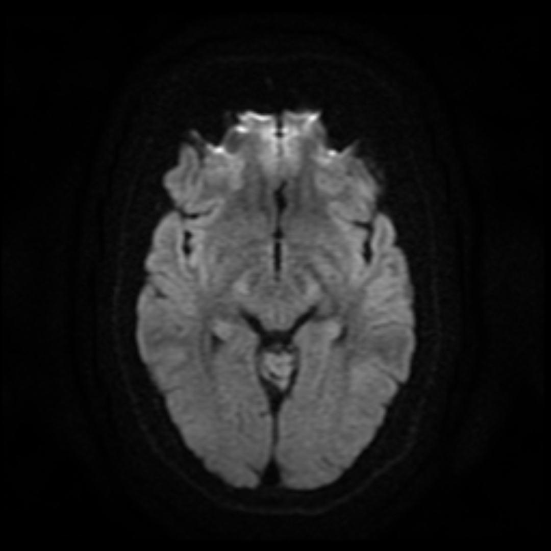
MRI brain is radiation-free and outperforms CT for small ischaemic strokes, multiple sclerosis, posterior fossa lesions, tumours, and dementia workup. It is the imaging of choice for chronic headache red flags, unexplained focal neurology, and suspected demyelination. Gadolinium contrast is added for tumour, infection, or MS relapse assessment.
MRI brain uses no ionising radiation and produces multiparametric images (T1, T2, FLAIR, DWI, SWI, MRA/MRV) that resolve tissue detail unmatched by CT. Diffusion-weighted imaging (DWI) shows acute ischaemia within minutes — sensitivity > 95% at < 24 h vs CT ~30%. FLAIR is best for MS plaques and gliosis. Susceptibility-weighted imaging (SWI) detects microhaemorrhages and cerebral amyloid angiopathy. Gadolinium contrast is added for suspected tumour, meningitis/encephalitis, MS relapse activity, and pituitary imaging. Gadolinium retention (nephrogenic systemic fibrosis) is a concern only in severe renal impairment (eGFR < 30). NICE and ACR "chronic headache" guidance: image only with red flags (thunderclap, new headache > 50, progressive, focal signs, immunosuppression, cancer history, wake from sleep). Incidental findings on brain MRI occur in ~2–3% and require judicious management (many are benign, e.g., arachnoid cysts, small meningiomas). MS diagnosis follows 2017 McDonald criteria — DIS (dissemination in space) and DIT (dissemination in time) demonstrable on MRI. Dementia workup: MRI helps exclude structural cause; regional atrophy patterns support Alzheimer, vascular, or frontotemporal aetiology. FDG-PET and amyloid/tau PET add molecular detail in specialist settings.
- Radiation
- None
- DWI for acute stroke
- Sensitivity > 95% at < 24 h
- Contrast (gadolinium)
- Tumour, infection, MS activity
- Incidental findings
- ~2–3% of asymptomatic scans
- MS criteria
- McDonald 2017 (DIS + DIT)
MRI brain sequences at a glance
| Sequence | What it shows |
|---|---|
| T1 | Anatomy; fat bright; CSF dark |
| T2 | Fluid, oedema, inflammation |
| FLAIR | MS plaques, gliosis (CSF suppressed) |
| DWI | Acute ischaemia, abscess |
| SWI | Microbleeds, calcification, iron |
| MRA | Non-contrast vessel imaging |
When MRI brain is justified
Chronic headache + any red flag (see ACR appropriateness).
Suspected MS or CIS (clinically isolated syndrome).
New focal neurology or progressive cognitive impairment.
Suspected pituitary lesion (± contrast).
Post-stroke workup (MRI-DWI within 24 h if diagnosis uncertain).
- New focal weakness or speech disturbance — stroke pathway.
- Papilloedema + headache — urgent MRI + venography (venous sinus thrombosis).
- Cancer history + new neurology — same-week MRI.
When brain imaging?
- 1Acute focal neurology?CT + CT angiography → MRI DWI if uncertain.
- 2Suspected MS?MRI brain + cervical cord with contrast.
- 3Chronic headache + red flags?MRI brain.
- 4Cognitive decline?MRI brain + relevant blood workup.
Related questions people ask
Frequently asked questions
- MRI outperforms CT for parenchymal and small-vessel disease.
- DWI is highly sensitive for acute stroke.
- MS diagnosis follows McDonald 2017 criteria.
- Contrast is not always required.
- Incidental findings are common — plan how to handle them.
References
2 sources- ACR Appropriateness CriteriaHeadache
US imaging appropriateness.
acr.org
- MAGNIMS/NICEMRI Criteria for MS Diagnosis
European MRI consortium.
magnims.eu
Related articles
 Imaging & Diagnostic Tests
Chest X-ray Explained: What It Shows, When You Need One, and Radiation Dose
A chest X-ray delivers a chest radiation dose equivalent to a few days of background radiation. Learn what it detects and what it misses.
 Imaging & Diagnostic Tests
CT Head Scan Explained: When It Is Needed for Stroke, Trauma and Headache
A non-contrast CT head is the first imaging test in stroke and head trauma. Learn NICE criteria, radiation dose, and what CT misses.
 Imaging & Diagnostic Tests
CTPA (CT Pulmonary Angiogram) Explained: Diagnosing Pulmonary Embolism
CTPA is the reference standard for pulmonary embolism. Learn indications, radiation dose, contrast risks, and pregnancy alternatives.