Bronchoscopy Explained: EBUS, BAL and Lung Biopsy
Flexible bronchoscopy inspects the airways and enables lavage, brushings and biopsies. Learn EBUS staging and TBLC in ILD.
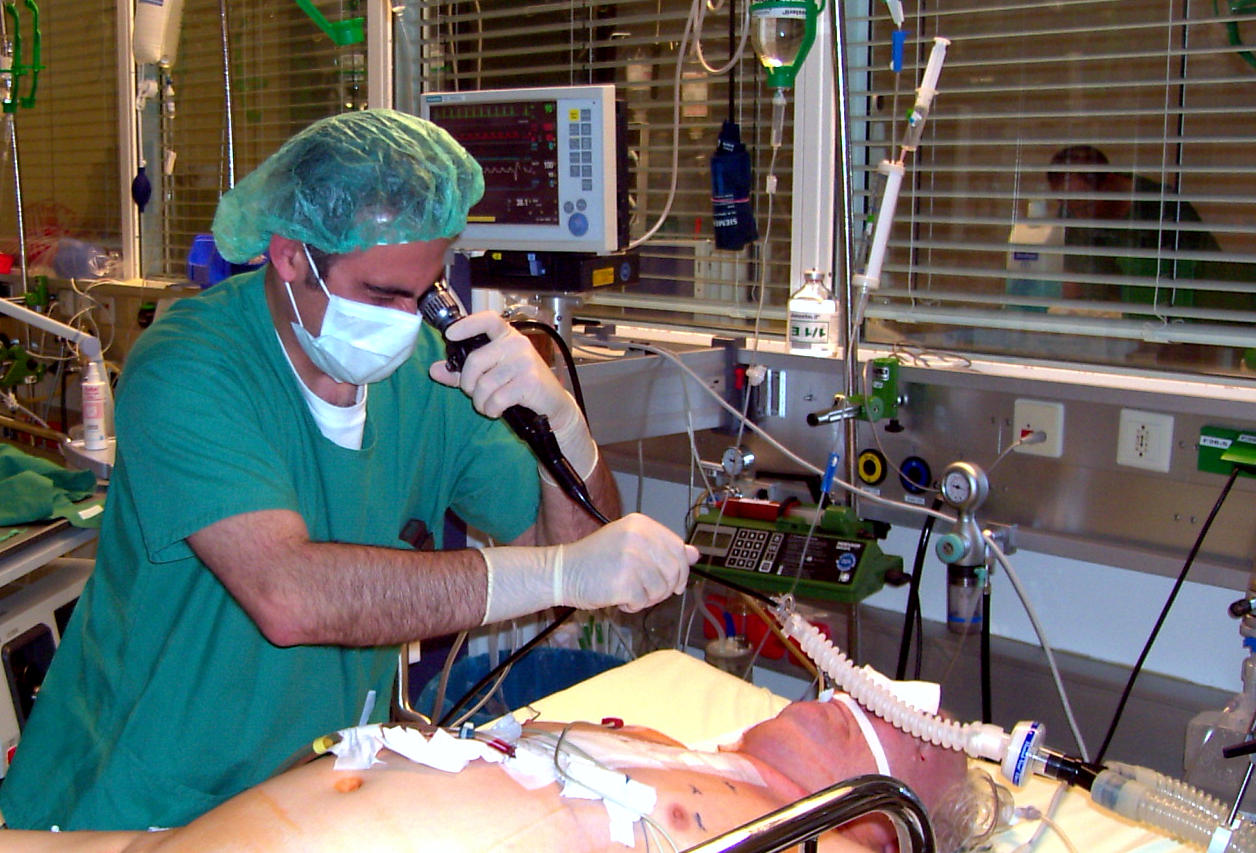
Flexible bronchoscopy passes a video scope through the nose or mouth into the airways, allowing inspection, bronchoalveolar lavage (BAL), brushings, biopsies, and interventions. EBUS-TBNA (endobronchial ultrasound + transbronchial needle aspiration) is the modern reference standard for mediastinal lymph-node staging in lung cancer.
Bronchoscopy is essential for many respiratory diagnoses. Standard indications: unexplained haemoptysis, endobronchial mass biopsy, ILD workup (BAL cellularity + transbronchial biopsy or cryobiopsy), removal of foreign body, therapeutic clearance of secretions, and lung cancer staging via EBUS-TBNA. EBUS-TBNA has replaced mediastinoscopy for most lymph-node staging in NSCLC — sensitivity > 90% for stations 2, 4, 7, 10, 11. Bronchoalveolar lavage (BAL) samples deep alveolar spaces: differential cellularity guides ILD diagnosis (neutrophilic in IPF, lymphocytic in HP or sarcoid, eosinophilic in EP). Transbronchial lung cryobiopsy (TBLC) is increasingly used in ILD as an alternative to surgical lung biopsy — larger tissue than forceps, moderate risk profile. Interventional bronchoscopy: endobronchial valve placement (LVR), rigid bronchoscopy for airway stenting or laser resection in central lesions. Sedation is conscious sedation or GA. Complications: minor bleeding (1–2%), pneumothorax after transbronchial biopsy (1–5%), rare cardiac/respiratory events. Preparation: fast 6 h, discuss anticoagulation.
- Sedation
- Conscious sedation or GA
- EBUS-TBNA
- Reference for mediastinal staging in NSCLC
- BAL cellularity
- Guides ILD diagnosis
- Cryobiopsy
- Alternative to surgical lung biopsy
- Pneumothorax risk (transbronchial biopsy)
- 1–5%
Bronchoscopy modalities
| Modality | When |
|---|---|
| Flexible white-light | Airway inspection |
| EBUS-TBNA | Mediastinal staging (NSCLC) |
| BAL | ILD workup, atypical infection |
| Transbronchial biopsy | Peripheral lesions, ILD |
| Cryobiopsy (TBLC) | ILD alternative to surgical biopsy |
When bronchoscopy is required
Unexplained haemoptysis.
Suspected central lung mass on CXR/CT.
Mediastinal lymphadenopathy of unclear cause.
ILD workup where HRCT is non-diagnostic.
Suspected atypical infection (TB, PJP, aspergillus).
- Massive haemoptysis — emergency bronchoscopy + IR embolisation.
- Central airway obstruction — rigid bronchoscopy + stent.
- Post-biopsy pneumothorax — chest drain if large or symptomatic.
Lung mass workup
- 1Central mass?Bronchoscopy + biopsy.
- 2Peripheral mass?CT-guided biopsy or navigational bronchoscopy.
- 3Mediastinal staging needed?EBUS-TBNA.
- 4ILD workup?BAL + cryobiopsy or surgical biopsy per MDT.
Related questions people ask
Frequently asked questions
- Bronchoscopy inspects and samples the airways.
- EBUS-TBNA is the modern staging standard in NSCLC.
- BAL differentiates ILDs and atypical infections.
- Cryobiopsy is a modern alternative to surgical biopsy.
- Pneumothorax is the main risk post-transbronchial biopsy.
References
2 sources- BTSFlexible Bronchoscopy Guidelines
British Thoracic Society.
brit-thoracic.org.uk
- ACCPDiagnosis and Management of Lung Cancer
American College of Chest Physicians.
journal.chestnet.org
Related articles
 Imaging & Diagnostic Tests
Chest X-ray Explained: What It Shows, When You Need One, and Radiation Dose
A chest X-ray delivers a chest radiation dose equivalent to a few days of background radiation. Learn what it detects and what it misses.
 Imaging & Diagnostic Tests
CT Head Scan Explained: When It Is Needed for Stroke, Trauma and Headache
A non-contrast CT head is the first imaging test in stroke and head trauma. Learn NICE criteria, radiation dose, and what CT misses.
 Imaging & Diagnostic Tests
CTPA (CT Pulmonary Angiogram) Explained: Diagnosing Pulmonary Embolism
CTPA is the reference standard for pulmonary embolism. Learn indications, radiation dose, contrast risks, and pregnancy alternatives.