HRCT Chest Explained: Interstitial Lung Disease, IPF and Bronchiectasis
HRCT chest is essential for diffuse lung disease. Learn UIP vs NSIP patterns and how HRCT drives IPF diagnosis.
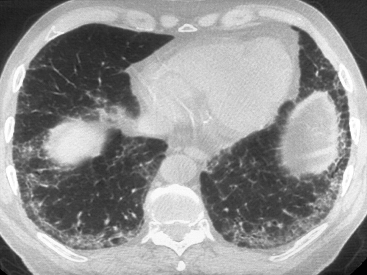
HRCT chest gives thin-slice, high-detail images of lung parenchyma — essential for interstitial lung disease (ILD), IPF diagnosis, and bronchiectasis. Radiation dose is ~5–8 mSv. A confident UIP pattern on HRCT alone diagnoses IPF (no biopsy needed) under 2022 ATS/ERS/JRS/ALAT criteria.
HRCT uses thin (0.5–1 mm) slice reconstruction and high spatial frequency algorithms to resolve fine lung architecture. Standard protocol: supine inspiratory + prone (to differentiate dependent atelectasis from early fibrosis) + expiratory (for air trapping). Radiation is comparable to standard chest CT (~5–8 mSv). ATS/ERS/JRS/ALAT 2022 IPF criteria: definite UIP pattern (subpleural, basal predominant honeycombing ± traction bronchiectasis) diagnoses IPF without biopsy; probable UIP or indeterminate patterns require MDT + possible surgical biopsy. Common ILD patterns: NSIP — ground-glass with basal reticulation, subpleural sparing (connective tissue disease); Hypersensitivity pneumonitis — mosaic attenuation + air trapping + centrilobular nodules; Sarcoid — perilymphatic nodules + upper-lobe distribution; Organising pneumonia — patchy consolidation, subpleural or peribronchial. Bronchiectasis: signet-ring sign (bronchus > accompanying artery), tram tracks, cystic dilation. HRCT drives MDT decisions and anti-fibrotic therapy in IPF (nintedanib, pirfenidone). Emerging: quantitative HRCT and AI progression scoring.
- Radiation
- ~5–8 mSv
- Best for
- ILD, IPF, bronchiectasis
- IPF diagnosis
- Definite UIP pattern alone (no biopsy needed)
- Standard sequences
- Supine + prone + expiratory
- ILD MDT
- Central to modern diagnosis
Key HRCT patterns + diagnoses
| Pattern | Suggests |
|---|---|
| Subpleural basal honeycombing | Definite UIP (IPF) |
| Ground-glass + basal reticulation, subpleural sparing | NSIP (CTD-ILD) |
| Mosaic attenuation + air trapping + centrilobular nodules | Hypersensitivity pneumonitis |
| Perilymphatic nodules, upper lobe | Sarcoidosis |
| Patchy consolidation, peripheral | Organising pneumonia |
| Signet-ring bronchi, tram tracks | Bronchiectasis |
When HRCT changes management
Unexplained persistent cough or breathlessness + abnormal CXR.
Suspected ILD on clinical grounds.
Baseline and follow-up in IPF on anti-fibrotic therapy.
Bronchiectasis assessment and phenotyping.
Occupational lung disease workup.
- Rapid progression of ILD — hospital admission for acute exacerbation.
- Persistent haemoptysis in bronchiectasis — bronchoscopy + BAE evaluation.
- New pulmonary hypertension in ILD — right-heart catheter.
ILD workup
- 1Suspected ILD?HRCT chest.
- 2Definite UIP?MDT confirms IPF; consider anti-fibrotic.
- 3Non-UIP pattern?MDT + serology + occupational history + biopsy.
- 4Bronchiectasis?HRCT + phenotype workup (CF gene, immune, aspergillus).
Related questions people ask
Frequently asked questions
- HRCT is the workhorse for diffuse lung disease.
- Definite UIP pattern diagnoses IPF without biopsy.
- Multiple ILD patterns are pattern-recognition based.
- Bronchiectasis is identified by classic morphology.
- MDT is central to ILD diagnosis.
References
2 sources- ATS/ERS/JRS/ALATIPF Diagnosis and Management (2022 update)
US/European/Japanese/Latin-American respiratory societies.
atsjournals.org
- BTSBronchiectasis Guidelines
British Thoracic Society.
brit-thoracic.org.uk
Related articles
 Imaging & Diagnostic Tests
Chest X-ray Explained: What It Shows, When You Need One, and Radiation Dose
A chest X-ray delivers a chest radiation dose equivalent to a few days of background radiation. Learn what it detects and what it misses.
 Imaging & Diagnostic Tests
CT Head Scan Explained: When It Is Needed for Stroke, Trauma and Headache
A non-contrast CT head is the first imaging test in stroke and head trauma. Learn NICE criteria, radiation dose, and what CT misses.
 Imaging & Diagnostic Tests
CTPA (CT Pulmonary Angiogram) Explained: Diagnosing Pulmonary Embolism
CTPA is the reference standard for pulmonary embolism. Learn indications, radiation dose, contrast risks, and pregnancy alternatives.