Spirometry Explained: FEV1, FVC, and Diagnosing COPD or Asthma
Spirometry is the reference test for airflow limitation. Learn FEV1/FVC ratios, reversibility, GOLD staging, and pitfalls.
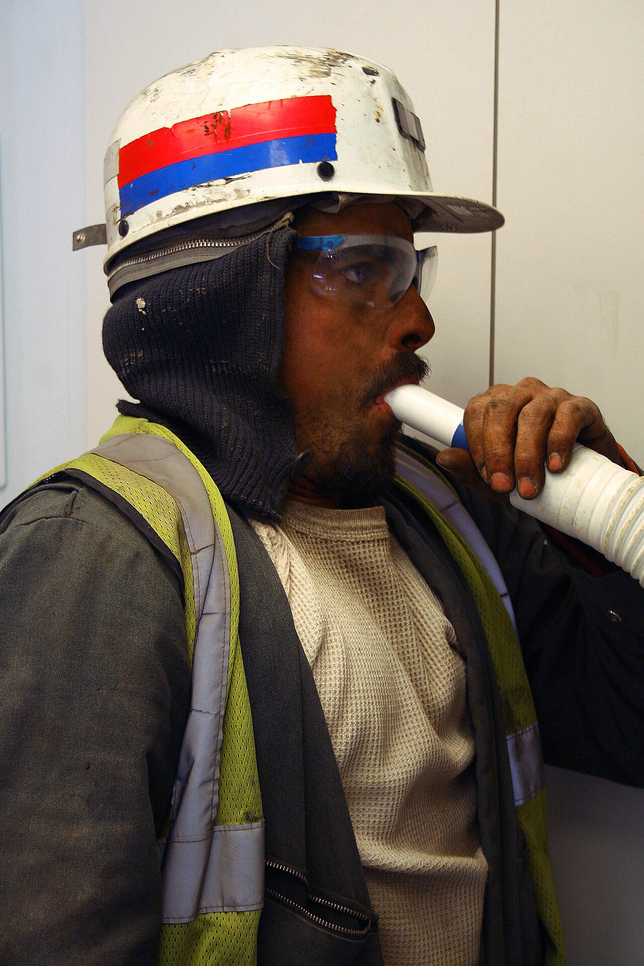
Spirometry measures forced expiratory volume in 1 second (FEV1) and forced vital capacity (FVC). Post-bronchodilator FEV1/FVC < 0.7 (or below the lower limit of normal) confirms airflow obstruction — diagnostic of COPD. In asthma, reversibility ≥ 12% AND ≥ 200 mL improvement in FEV1 after bronchodilator supports diagnosis. NICE NG115 (2018) requires spirometry for COPD diagnosis; NICE NG80 (asthma, 2024 update) endorses spirometry + FeNO + PEF variability.
Spirometry is the reference physiological test for airflow limitation. GOLD (Global Initiative for COPD, 2024) uses FEV1 percentage predicted for severity: GOLD 1 ≥ 80% (mild), 2 = 50–79% (moderate), 3 = 30–49% (severe), 4 < 30% (very severe). ATS/ERS 2019 standardisation improved quality: at least three acceptable manoeuvres, two reproducible within 150 mL, and back-extrapolated volume < 5%. Use of race-neutral reference equations (GLI 2022 global) is replacing race-specific equations — controversial but evolving. Asthma: reversibility test with 400 mcg salbutamol via spacer; positive = ≥ 12% AND ≥ 200 mL rise in FEV1 (adult NICE criterion). Additional testing where uncertain: FeNO (exhaled nitric oxide) for eosinophilic airway inflammation, methacholine or mannitol challenge for bronchial hyperresponsiveness, and PEF diary. Peak expiratory flow (PEF) diary variability > 20% supports asthma. Restrictive pattern (low FEV1, low FVC, normal ratio) suggests interstitial lung disease, obesity, neuromuscular disease — needs lung volumes and gas transfer to confirm.
- Airflow obstruction
- Post-bronchodilator FEV1/FVC < 0.7 (or below LLN)
- Asthma reversibility
- ≥ 12% AND ≥ 200 mL FEV1 improvement
- COPD severity
- GOLD 1–4 by FEV1 % predicted
- Quality standard
- ATS/ERS 2019
- Reference equations
- GLI 2022 global
Interpreting spirometry patterns
| Pattern | FEV1/FVC | FVC | Common causes |
|---|---|---|---|
| Obstructive | < 0.7 | Normal or low | COPD, asthma, bronchiectasis |
| Restrictive | ≥ 0.7 | Low | ILD, obesity, kyphosis, neuromuscular |
| Mixed | < 0.7 | Low | Combined obstruction + restriction |
| Normal | ≥ 0.7 | Normal | No airflow limitation |
Preparation
No smoking 1 hour before.
No short-acting bronchodilator 4 h; long-acting 12–24 h (unless doing reversibility).
No large meal 2 h; avoid tight clothing.
Bring current inhalers.
Post-viral: wait 2–4 weeks after respiratory infection.
- Severe airflow obstruction FEV1 < 30% predicted — respiratory referral.
- Rapid decline in FEV1 — investigate exacerbation drivers.
- Restrictive pattern with breathlessness — HRCT and gas transfer.
Airflow obstruction workup
- 1Post-BD FEV1/FVC < 0.7?Airflow obstruction — likely COPD if smoker.
- 2Reversibility ≥ 12 % + ≥ 200 mL?Supports asthma.
- 3Restrictive pattern?Full lung volumes + gas transfer.
- 4Uncertain?FeNO + PEF diary + challenge test.
Related questions people ask
Frequently asked questions
- Spirometry is the reference test for airflow limitation.
- FEV1/FVC < 0.7 post-BD confirms obstruction.
- Reversibility supports asthma.
- GOLD stages COPD severity.
- Follow ATS/ERS 2019 quality standards.
References
2 sources- GOLD 2024 ReportCOPD
Global initiative for COPD.
goldcopd.org
- ATS/ERS 2019Standardisation of Spirometry
US/European respiratory societies.
atsjournals.org
Related articles
 Imaging & Diagnostic Tests
Chest X-ray Explained: What It Shows, When You Need One, and Radiation Dose
A chest X-ray delivers a chest radiation dose equivalent to a few days of background radiation. Learn what it detects and what it misses.
 Imaging & Diagnostic Tests
CT Head Scan Explained: When It Is Needed for Stroke, Trauma and Headache
A non-contrast CT head is the first imaging test in stroke and head trauma. Learn NICE criteria, radiation dose, and what CT misses.
 Imaging & Diagnostic Tests
CTPA (CT Pulmonary Angiogram) Explained: Diagnosing Pulmonary Embolism
CTPA is the reference standard for pulmonary embolism. Learn indications, radiation dose, contrast risks, and pregnancy alternatives.