12-Lead ECG Explained: What It Shows, Common Abnormalities and Limits
The ECG is fast, cheap, and central to cardiology. Learn what it captures, common patterns, and when a normal ECG is not reassuring.
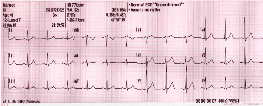
The 12-lead ECG records electrical activity through 10 electrodes producing 12 views. It is essential for chest pain, palpitations, breathlessness, syncope, and pre-operative assessment. A normal ECG does NOT exclude coronary disease, PE, or aortic dissection.
The 12-lead ECG is one of the most commonly performed clinical investigations. Reading follows a systematic sequence: rate, rhythm, axis, P wave, PR interval, QRS morphology, ST segment, T wave, QT interval. Key patterns: STEMI (ST elevation ≥ 1 mm in ≥ 2 contiguous limb leads or ≥ 2 mm in ≥ 2 contiguous chest leads) — immediate primary PCI or thrombolysis. New LBBB with chest pain is treated like STEMI on modern protocols (Sgarbossa criteria refine ischaemia recognition in known LBBB). NSTEMI shows ST depression or T-wave inversion + raised troponin. AF: irregularly irregular rhythm, absent P waves — CHA₂DS₂-VASc guides anticoagulation. Long QT (QTc > 500 ms) increases torsades risk. Hyperkalaemia progresses through peaked T waves → PR prolongation → wide QRS → sine wave. Digoxin causes "reverse tick" ST depression. Limitations: single ECG snapshots miss paroxysmal arrhythmia — use ambulatory monitoring (Holter, event recorder, implantable loop recorder). AI ECG interpretation is a rapidly evolving field but human overread remains standard.
- Normal PR
- 120 – 200 ms
- Normal QRS
- < 120 ms
- Corrected QT (QTc)
- M < 440, F < 460 ms
- STEMI
- ST elevation ≥ 1 mm limb / ≥ 2 mm chest
- AF
- Irregularly irregular, absent P waves
Common ECG patterns
| Pattern | Meaning |
|---|---|
| Peaked symmetric T waves | Hyperkalaemia |
| ST elevation ≥ 1 mm in ≥ 2 contiguous leads | STEMI |
| Deep symmetric T-wave inversion (Wellens) | Proximal LAD stenosis — high risk |
| S1Q3T3 | Classic PE pattern (uncommon) |
| Delta wave + short PR | WPW pre-excitation |
When ECG is essential
Any chest pain — immediate ECG (within 10 minutes for suspected ACS).
Palpitations, dizziness, or syncope.
Newly diagnosed heart failure or persistent dyspnoea.
Pre-operative screening in patients > 65 or with cardiovascular disease.
Assessment before initiating QT-prolonging drugs.
- Chest pain + ST elevation — primary PCI within 90 minutes.
- Wide-complex tachycardia — assume VT until proven otherwise.
- QTc > 500 ms + syncope or arrhythmia symptoms — urgent cardiology.
Palpitations workup
- 1ECG shows AF/flutter?Rate control + CHA₂DS₂-VASc anticoagulation.
- 2ECG normal but symptoms recurrent?Ambulatory monitor (24–72 h Holter, patch, event recorder).
- 3Suspected SVT / pre-excitation?Cardiology + electrophysiology referral.
Related questions people ask
Frequently asked questions
- ECG is fast, cheap, and universally available.
- Never rule out cardiac disease on a normal single ECG.
- STEMI = immediate primary PCI pathway.
- AF is detected by irregular rhythm + absent P waves.
- Ambulatory monitors extend the reach of the 12-lead ECG.
References
2 sources- ESC 2023 ACS Guidelines
European Society of Cardiology.
academic.oup.com
- AHARecommendations for the Standardization and Interpretation of the ECG
AHA standard.
ahajournals.org
Related articles
 Imaging & Diagnostic Tests
Chest X-ray Explained: What It Shows, When You Need One, and Radiation Dose
A chest X-ray delivers a chest radiation dose equivalent to a few days of background radiation. Learn what it detects and what it misses.
 Imaging & Diagnostic Tests
CT Head Scan Explained: When It Is Needed for Stroke, Trauma and Headache
A non-contrast CT head is the first imaging test in stroke and head trauma. Learn NICE criteria, radiation dose, and what CT misses.
 Imaging & Diagnostic Tests
CTPA (CT Pulmonary Angiogram) Explained: Diagnosing Pulmonary Embolism
CTPA is the reference standard for pulmonary embolism. Learn indications, radiation dose, contrast risks, and pregnancy alternatives.