Echocardiogram Explained: Ejection Fraction, Valves and Heart Function
Echo is the workhorse for heart function. Learn ejection fraction ranges, HFpEF vs HFrEF, valve assessment, and TOE vs TTE.
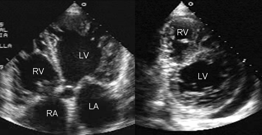
A transthoracic echocardiogram (TTE) uses ultrasound to assess heart chambers, valves, and function. Normal left ventricular ejection fraction (LVEF) is 55–70%. Heart failure is classified as reduced EF (HFrEF, ≤ 40%), mildly reduced (HFmrEF, 41–49%), or preserved (HFpEF, ≥ 50%). Trans-oesophageal echo (TOE) gives better valve/atrial detail and is used for endocarditis, atrial thrombus, or intraoperative assessment.
The echocardiogram is central to modern cardiology because it is radiation-free, portable, and gives real-time functional data. ESC 2021 heart-failure guideline classifies LVEF: HFrEF ≤ 40% (all four foundation therapies apply — ACEi/ARNI + beta-blocker + MRA + SGLT2i), HFmrEF 41–49% (foundation therapies increasingly recommended), HFpEF ≥ 50% (SGLT2i now class I recommendation on EMPEROR-Preserved and DELIVER data). Valvular grading follows AHA/ACC 2020 (aortic stenosis peak velocity ≥ 4 m/s or mean gradient ≥ 40 mmHg is severe; mitral regurgitation grading by regurgitant volume, effective regurgitant orifice area, PISA). Diastolic dysfunction: E/e′ ratio > 14, left atrial volume index > 34 mL/m², tricuspid regurgitation velocity > 2.8 m/s indicate impairment. TOE indications: endocarditis, atrial thrombus (before cardioversion), aortic dissection, prosthetic valve assessment, intraoperative. Stress echo (dobutamine or exercise) assesses inducible ischaemia or valve behaviour under load.
- Radiation
- None
- Normal EF
- 55 – 70%
- HFrEF
- EF ≤ 40%
- HFpEF
- EF ≥ 50% + evidence of diastolic dysfunction
- TOE indications
- Endocarditis, atrial thrombus, dissection
Ejection fraction categories (ESC 2021)
| LVEF | Category | Treatment focus |
|---|---|---|
| ≤ 40% | HFrEF | ARNI/ACEi + BB + MRA + SGLT2i |
| 41 – 49% | HFmrEF | Foundation therapies (mostly transferable) |
| ≥ 50% | HFpEF | SGLT2i (class I), diuretics, comorbidity focus |
Understanding an echo report
LV dimensions: internal diastolic diameter (LVIDd) and wall thicknesses; dilated LV with wall thinning suggests dilated cardiomyopathy.
Regional wall motion: hypokinesia in a coronary territory suggests infarction.
Diastolic function: E/A ratio, E/e′, LA volume index, TR velocity.
Valves: peak/mean gradients + orifice area + regurgitant severity.
Right ventricle: TAPSE (< 17 mm impaired), RV S′ velocity, RV dilation.
- Severe aortic stenosis + symptoms — surgical or TAVI referral.
- EF ≤ 35% + wide QRS or symptomatic — ICD/CRT assessment.
- Suspected endocarditis + murmur — urgent echo (TTE then TOE).
Suspected heart failure workup
- 1Breathless + oedema?BNP + NT-proBNP.
- 2BNP raised or high probability?TTE within 2 weeks (NICE).
- 3HFrEF confirmed?Foundation therapies + specialist referral.
- 4Uncertain aetiology?Cardiac MRI or coronary angiography.
Related questions people ask
Frequently asked questions
- Echo is the workhorse of heart function assessment.
- HFrEF (≤ 40%) is treated with 4 foundation therapies.
- HFpEF now has SGLT2i class I recommendation.
- TOE is reserved for endocarditis, atrial thrombus, dissection.
- Interpret with clinical context — imaging alone is never enough.
References
2 sources- ESC 2021 Heart Failure Guideline (with 2023 focused update)
European Society of Cardiology.
academic.oup.com
- ACC/AHA 2020 Valvular Heart Disease Guideline
US cardiology.
jacc.org
Related articles
 Imaging & Diagnostic Tests
Chest X-ray Explained: What It Shows, When You Need One, and Radiation Dose
A chest X-ray delivers a chest radiation dose equivalent to a few days of background radiation. Learn what it detects and what it misses.
 Imaging & Diagnostic Tests
CT Head Scan Explained: When It Is Needed for Stroke, Trauma and Headache
A non-contrast CT head is the first imaging test in stroke and head trauma. Learn NICE criteria, radiation dose, and what CT misses.
 Imaging & Diagnostic Tests
CTPA (CT Pulmonary Angiogram) Explained: Diagnosing Pulmonary Embolism
CTPA is the reference standard for pulmonary embolism. Learn indications, radiation dose, contrast risks, and pregnancy alternatives.