Potassium Blood Test: Hyperkalaemia, Hypokalaemia and Pseudo-Hyperkalaemia Explained
Potassium > 6.5 is a medical emergency. Learn ECG changes, spurious elevation, and safe correction.
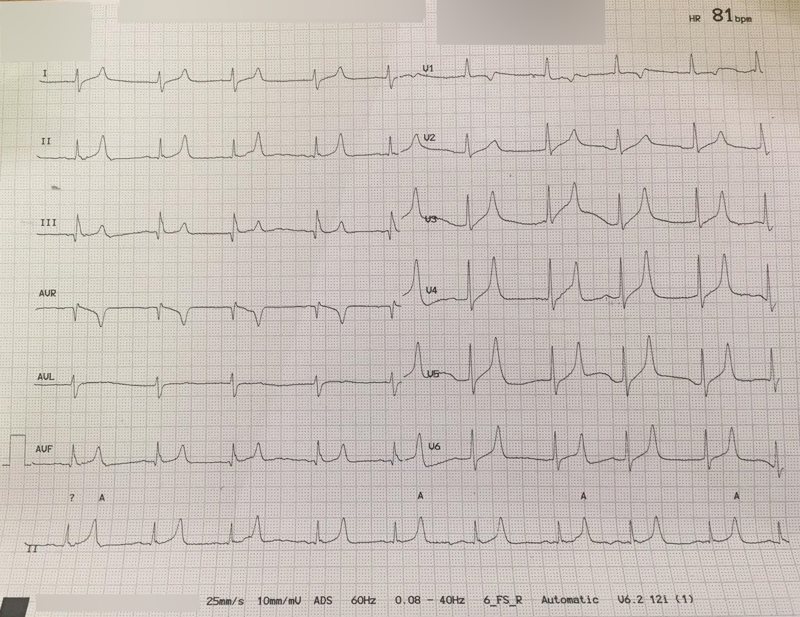
Normal potassium is 3.5–5.0 mmol/L. Above 6.5 mmol/L or any level with ECG changes is a medical emergency. Rule out pseudo-hyperkalaemia (haemolysis, delayed processing, tight tourniquet) with a repeat sample. Treatment sequence: calcium gluconate → insulin + glucose → salbutamol → definitive removal (dialysis or new binders).
Hyperkalaemia is the electrolyte disturbance with the highest immediate mortality risk. The 2023 UK Renal Association / RA guideline defines severe hyperkalaemia as ≥ 6.5 mmol/L or ≥ 6.0 with ECG changes; treatment must not wait for the confirmatory sample. ECG changes progress: tall tented T waves → PR prolongation → wide QRS → sine-wave → asystole. Standard emergency sequence: (1) IV calcium gluconate 10% 10 mL to stabilise myocardium (does not lower potassium); (2) insulin 10 units in 25 g glucose IV to shift potassium intracellularly; (3) nebulised salbutamol 10–20 mg for additive shift; (4) definitive removal — dialysis for oliguric AKI, patiromer or sodium zirconium cyclosilicate for chronic RAAS-inhibitor-associated hyperkalaemia. Pseudo-hyperkalaemia (haemolysis, delayed lab processing, thrombocytosis, tight tourniquet) is common — always repeat if borderline and unexpected. Hypokalaemia (< 3.5) is usually GI losses, diuretics, or hyperaldosteronism; check magnesium (low Mg blocks potassium repletion) and correct together.
- Normal K+
- 3.5 – 5.0 mmol/L
- Emergency
- ≥ 6.5 or ECG changes
- First drug
- Calcium gluconate (myocardial stabilisation)
- Shifter
- Insulin 10 U + 25 g glucose
- Remover
- Dialysis, patiromer, sodium zirconium cyclosilicate
Hyperkalaemia severity + management
| K+ (mmol/L) | Severity | Action |
|---|---|---|
| 5.1 – 5.9 | Mild | Review K+ intake, drugs; recheck. |
| 6.0 – 6.4 | Moderate | ECG. Stop offenders. Insulin + glucose if ECG changes. |
| ≥ 6.5 | Severe | Emergency. Full sequence: Ca → insulin → salbutamol → removal. |
Pseudo-hyperkalaemia — always consider
Haemolysed sample: tight tourniquet, small needle, delayed transport.
Extreme thrombocytosis (platelets > 1000 × 10⁹/L): potassium released during clotting.
Extreme leukocytosis in CLL.
Delayed processing: send to lab within 1 hour and process promptly.
- Any K+ ≥ 6.5 mmol/L, or ≥ 6.0 with ECG changes — emergency.
- Weakness or palpitations + K+ ≥ 6.0 — immediate ECG.
- On ACE inhibitor / spironolactone with rising K+ — reassess drug + kidney function.
Hyperkalaemia workup
- 1Any ECG changes?IV calcium gluconate immediately.
- 2K+ ≥ 6.0?Insulin + glucose. Nebulised salbutamol. Stop RAAS blockers.
- 3Anuric AKI or refractory?Dialysis.
- 4Chronic, mild, on essential RAAS inhibitor?Patiromer or sodium zirconium cyclosilicate.
Related questions people ask
Frequently asked questions
- K+ ≥ 6.5 or ECG changes = emergency.
- Calcium first (myocardial protection).
- Insulin + glucose shifts potassium.
- Dialysis or new binders remove potassium.
- Always exclude pseudo-hyperkalaemia.
References
2 sources- UK Kidney AssociationHyperkalaemia Guideline (2023)
Emergency management.
ukkidney.org
- European Resuscitation CouncilCardiac Arrest in Special Circumstances
Emergency guidance.
erc.edu
Evidence Snapshot
This snapshot shows verified evidence records matched to this article. It is not a diagnosis or personal medical advice.
Serum magnesium reflects only a small fraction of total body magnesium, most of which sits inside cells and bone, so a normal serum magnesium value does not by itself exclude tissue-level magnesium depletion.
Limitation. Tests that better reflect intracellular magnesium exist but are not part of routine panels and may not change routine practice.
Related articles
 Laboratory Tests
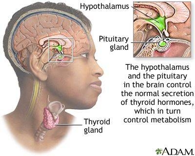
TSH Test: Normal Range, High, Low & What Your Result Actually Means
TSH is the single most important thyroid number. Understand normal ranges by age and pregnancy, what a high or low value really tells you, and the two follow-up tests that clinch the diagnosis.
 Laboratory Tests
HbA1c Test: Normal Range, Prediabetic, Diabetic Cut-offs & How to Lower It
HbA1c reflects your average blood sugar over ~3 months. Understand every threshold — normal, prediabetes, diabetes — and the realistic actions that move the number.
 Laboratory Tests
LDL Cholesterol: Optimal Numbers, High Numbers, and How to Read Yours
LDL is the "bad" cholesterol only if you leave the story there. Understand optimal targets by risk group, the ApoB perspective, and the interventions that actually shift the number.