Capsule Endoscopy Explained: Investigating Obscure GI Bleeding
A pill camera captures the small bowel invisible to OGD and colonoscopy. Learn indications, retention risk and complementary tests.
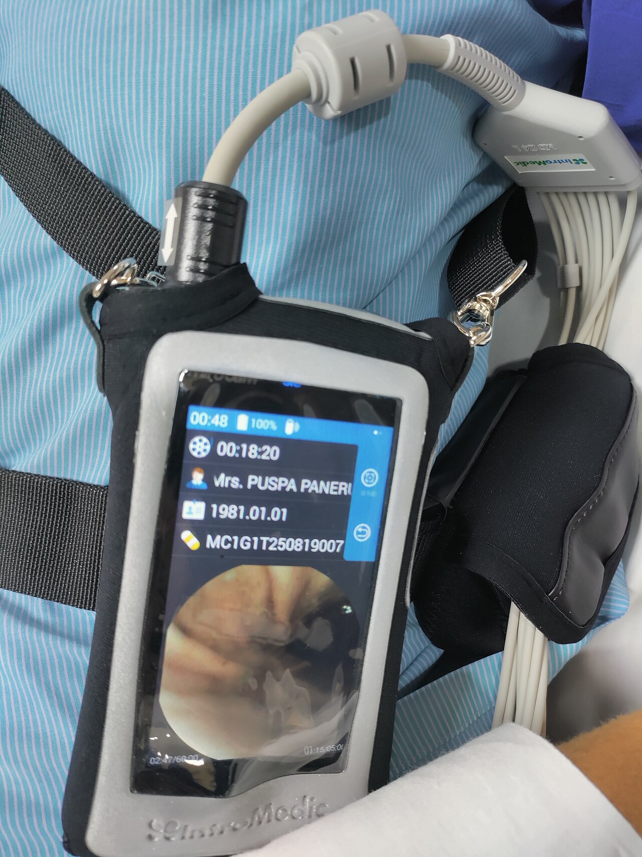
Video capsule endoscopy (VCE) is a swallowed pill camera that images the small bowel — invisible to OGD and colonoscopy. First-line for obscure GI bleeding or iron-deficiency anaemia after normal OGD + colonoscopy. Retention risk 1–2%; a patency capsule may be used if stricture is suspected.
Capsule endoscopy transformed small-bowel investigation. Standard indications: obscure GI bleeding (bleeding continues after normal OGD + colonoscopy), iron-deficiency anaemia with negative bidirectional endoscopy, suspected Crohn disease with normal ileocolonoscopy, coeliac disease complications (ulcerative jejunoileitis, small-bowel lymphoma), polyposis surveillance (Peutz-Jeghers, familial adenomatous polyposis after duodenoscopy). Modern capsules (Pillcam SB3, MiroCam, Olympus EndoCapsule) transmit ~8–12 hours of imaging. Findings: angiodysplasia (commonest cause of obscure bleeding in older adults), Crohn ulceration, small-bowel tumours (rare but important), coeliac villous atrophy. Limitations: cannot biopsy, cannot control view/direction, and cannot be used if intestinal obstruction/stricture is suspected without prior patency capsule. NICE, BSG, ESGE endorse capsule endoscopy as first-line small-bowel imaging in obscure bleeding — usually preceded by CT abdomen if massive bleeding. Colon capsule endoscopy is emerging for incomplete colonoscopy or contraindications to conventional colonoscopy — accepted by NICE 2020 draft guidance.
- Purpose
- Small-bowel imaging beyond OGD/colonoscopy
- Duration
- ~8–12 h recording
- Retention risk
- 1–2%
- Stricture risk?
- Patency capsule first
- First-line for
- Obscure GI bleeding, IDA after negative endoscopy
Small-bowel test choices
| Question | Best test |
|---|---|
| Obscure GI bleeding | Capsule endoscopy |
| Suspected Crohn stricturing | MR enterography |
| Small-bowel tumour | Capsule + CT/MR enterography |
| Coeliac complication | Capsule + biopsy via deep enteroscopy |
Preparation
Clear-fluid diet 24 h before; fast 8 h before.
Small-bowel bowel prep (varies by service) improves image quality.
Simethicone reduces bubbles.
Avoid magnets and MRI until capsule confirmed passed.
Return recorder after transit; capsule passed in stool over days.
- Abdominal pain + distension after capsule — suspect retention.
- No confirmation of passage after 2 weeks — imaging.
- Ongoing bleeding despite negative capsule — deep enteroscopy or CT angiography.
Obscure GI bleeding workup
- 1OGD + colonoscopy negative?Capsule endoscopy.
- 2Capsule positive?Deep enteroscopy (single- or double-balloon) for biopsy/treatment.
- 3Stricture suspected?Patency capsule or MR enterography first.
Related questions people ask
Frequently asked questions
- Capsule endoscopy is first-line for obscure GI bleeding.
- It images the small bowel that OGD and colonoscopy miss.
- Retention risk 1–2% — patency capsule mitigates.
- Cannot biopsy — plan deep enteroscopy after positive findings.
- Emerging role for colon capsule endoscopy.
References
2 sources- BSGSmall-bowel endoscopy guidelines
British Society of Gastroenterology.
bsg.org.uk
- ESGESmall-bowel Endoscopy Guidelines
European endoscopy society.
esge.com
Related articles
 Imaging & Diagnostic Tests
Chest X-ray Explained: What It Shows, When You Need One, and Radiation Dose
A chest X-ray delivers a chest radiation dose equivalent to a few days of background radiation. Learn what it detects and what it misses.
 Imaging & Diagnostic Tests
CT Head Scan Explained: When It Is Needed for Stroke, Trauma and Headache
A non-contrast CT head is the first imaging test in stroke and head trauma. Learn NICE criteria, radiation dose, and what CT misses.
 Imaging & Diagnostic Tests
CTPA (CT Pulmonary Angiogram) Explained: Diagnosing Pulmonary Embolism
CTPA is the reference standard for pulmonary embolism. Learn indications, radiation dose, contrast risks, and pregnancy alternatives.