Upper GI Endoscopy (OGD) Explained: Dyspepsia, Barrett's and Coeliac Biopsy
OGD directly visualises the oesophagus, stomach and duodenum. Learn NICE red-flag criteria, Barrett's surveillance, and coeliac biopsy sites.
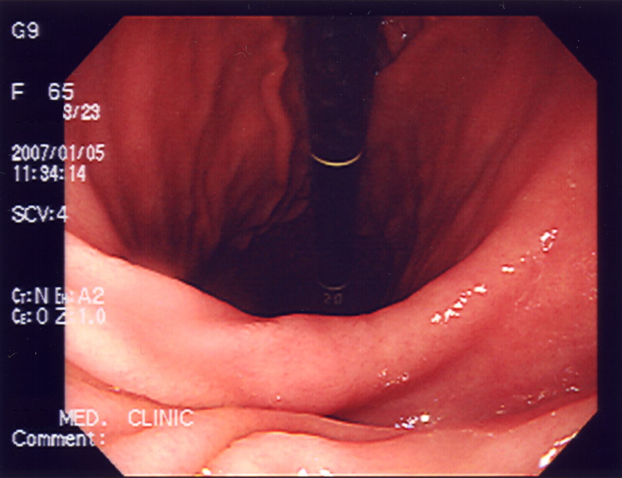
OGD (also called EGD or gastroscopy) inspects the oesophagus, stomach and duodenum through a flexible camera. NICE recommends urgent (2-week) OGD for age ≥ 55 with weight loss and any of: upper abdominal pain, reflux, dyspepsia — plus dysphagia at any age, or upper GI bleeding. Biopsies confirm H. pylori, Barrett's, coeliac disease, and cancer.
OGD (upper GI endoscopy) is the reference standard for upper GI symptoms. NICE NG12 recommends urgent 2-week wait referral for: any-age dysphagia; upper GI bleeding; age ≥ 55 with weight loss + upper abdominal pain, reflux, or dyspepsia. In dyspepsia without red flags, "test-and-treat" for H. pylori (stool antigen or urea breath test) is first-line; endoscopy is reserved for refractory symptoms or red flags. Barrett's oesophagus (BSG 2014, updated 2019) surveillance depends on segment length, dysplasia, and patient factors: 3–5 years for short-segment non-dysplastic; annual or endotherapy for dysplasia. Coeliac disease is confirmed with duodenal biopsies (≥ 4 from second part + 1–2 from bulb) — never diagnose coeliac on serology alone in adults. Modern therapeutic endoscopy: variceal banding, ulcer haemostasis, and endoscopic submucosal dissection (ESD) for early gastric/oesophageal cancer.
- Preparation
- Fast 6 hours (2 hours clear fluids)
- Sedation
- Optional — throat spray or IV sedation
- H. pylori test
- Rapid urease + histology
- Coeliac biopsies
- ≥ 4 from D2 + 1–2 from bulb
- Perforation risk
- ~1 in 3000
When OGD is indicated (NICE NG12)
| Presentation | Referral |
|---|---|
| Any-age dysphagia | Urgent 2-WW OGD |
| Upper GI bleeding (haematemesis/melaena) | Same-day admission |
| ≥ 55 y weight loss + reflux/dyspepsia/pain | Urgent 2-WW OGD |
| Dyspepsia without red flags | Test-and-treat H. pylori first |
| Refractory reflux on PPI | Elective OGD |
Barrett's oesophagus surveillance (BSG)
Short-segment (< 3 cm) non-dysplastic Barrett's: 3–5 year surveillance.
Long-segment (≥ 3 cm) non-dysplastic Barrett's: 2–3 year surveillance.
Low-grade dysplasia: repeat OGD 6 months; endoscopic eradication now preferred.
High-grade dysplasia or intramucosal cancer: endoscopic submucosal resection + radiofrequency ablation.
- Vomiting blood or coffee-ground vomiting — emergency admission.
- Melaena (black tarry stools) — same-day OGD.
- Progressive dysphagia — urgent OGD.
Dyspepsia pathway
- 1Any red flag?Urgent OGD.
- 2Refractory dyspepsia?Test-and-treat H. pylori, then OGD if failed.
- 3Iron-deficiency anaemia + GI symptoms?OGD + colonoscopy.
- 4Post-Barrett's?BSG surveillance interval.
Related questions people ask
Frequently asked questions
- OGD is the reference standard for upper GI symptoms.
- NICE red flags trigger urgent 2-week referral.
- H. pylori test-and-treat comes before OGD in uncomplicated dyspepsia.
- Coeliac disease needs duodenal biopsies.
- Barrett's surveillance intervals depend on length and dysplasia.
References
2 sources- NICE NG12Suspected cancer: recognition and referral
UK national guideline.
nice.org.uk
- BSGBarrett's Oesophagus Guidelines
British Society of Gastroenterology.
bsg.org.uk
Related articles
 Imaging & Diagnostic Tests
Chest X-ray Explained: What It Shows, When You Need One, and Radiation Dose
A chest X-ray delivers a chest radiation dose equivalent to a few days of background radiation. Learn what it detects and what it misses.
 Imaging & Diagnostic Tests
CT Head Scan Explained: When It Is Needed for Stroke, Trauma and Headache
A non-contrast CT head is the first imaging test in stroke and head trauma. Learn NICE criteria, radiation dose, and what CT misses.
 Imaging & Diagnostic Tests
CTPA (CT Pulmonary Angiogram) Explained: Diagnosing Pulmonary Embolism
CTPA is the reference standard for pulmonary embolism. Learn indications, radiation dose, contrast risks, and pregnancy alternatives.