Carotid Doppler Ultrasound Explained: TIA Workup and Stenosis Grading
Carotid ultrasound is the first vascular test after TIA or stroke. Learn NASCET stenosis grading and when surgery reduces stroke risk.
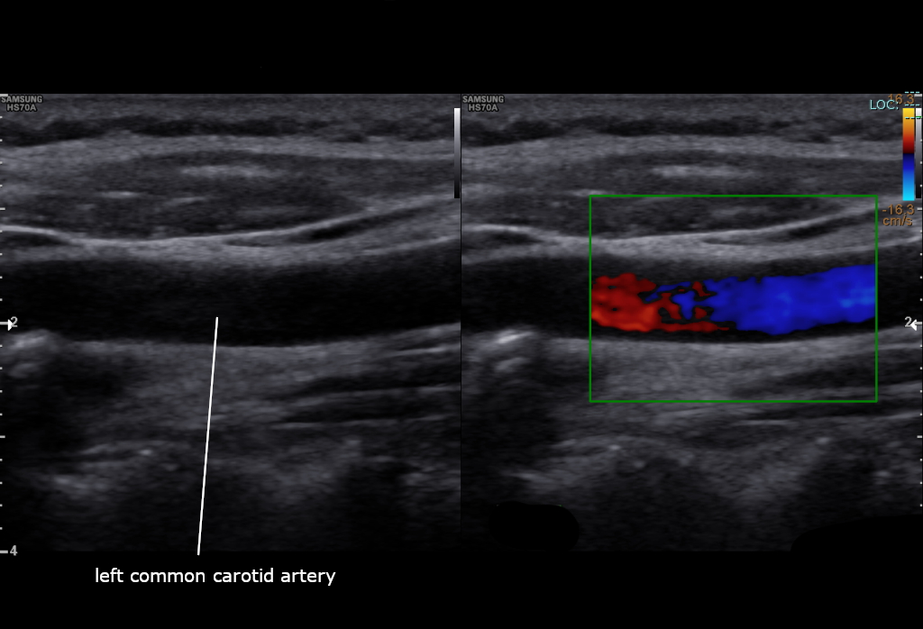
Carotid Doppler ultrasound is the first-line vascular test after TIA or minor stroke, measuring stenosis by peak systolic velocity (PSV) and end-diastolic velocity (EDV). NICE recommends symptomatic patients with > 50% NASCET stenosis be considered for carotid endarterectomy or stenting within 14 days. Asymptomatic stenosis is generally managed medically unless > 70% in fit patients.
Carotid Doppler combines B-mode imaging and pulsed-wave Doppler to quantify carotid stenosis non-invasively without radiation or contrast. Peak systolic velocity (PSV) is the primary metric: > 125 cm/s suggests ≥ 50% NASCET stenosis; > 230 cm/s suggests ≥ 70%; > 400 cm/s + very high EDV suggests near-occlusion. Confirmatory CT or MR angiography is standard before intervention. NICE NG128 and ESO 2021 recommend: symptomatic TIA/stroke patients with 50–99% carotid stenosis should be considered for carotid endarterectomy (CEA) or stenting within 14 days of symptom onset — the earlier the greater benefit (NNT ~5 for stroke prevention in the first 2 weeks). Beyond 6 months, benefit is minimal. Asymptomatic > 70% stenosis: intervention is controversial in the modern era of intensive medical therapy (statin + antiplatelet + BP control) — most patients are managed medically unless multiple lesion or contralateral occlusion. Carotid intima-media thickness (CIMT) is a research/screening measure, not routinely used clinically. Rare vertebral artery Doppler is added when posterior circulation events are suspected.
- Radiation
- None
- ≥ 50% stenosis PSV
- > 125 cm/s
- ≥ 70% stenosis PSV
- > 230 cm/s
- Symptomatic urgent surgery window
- 14 days
- Asymptomatic management
- Intensive medical therapy
Carotid stenosis + intervention
| Stenosis | Symptomatic | Asymptomatic |
|---|---|---|
| < 50% | Medical | Medical |
| 50–69% | Consider CEA/CAS (some benefit) | Medical |
| 70–99% | CEA/CAS within 14 days (strong benefit) | Consider — controversial |
| 100% occluded | Medical (revascularisation not helpful) | Medical |
When to order carotid Doppler
After TIA or minor stroke in the anterior circulation — urgent scan.
Bruit + high vascular risk profile (occasionally).
Follow-up after carotid endarterectomy or stenting.
NOT for asymptomatic screening in low-risk patients (USPSTF grade D).
- TIA or minor stroke — TIA clinic + carotid Doppler within 24 h.
- Fluctuating or crescendo TIAs — hospital admission.
- Doppler ≥ 70% stenosis symptomatic side — vascular surgery within 14 days.
Post-TIA workup
- 1TIA or minor stroke?ABCD² + carotid Doppler + ECG/72-h monitor + brain imaging.
- 2Carotid ≥ 50% ipsilateral to event?CT or MR angiography confirmation → CEA/CAS < 14 days.
- 3AF detected?Anticoagulation.
Related questions people ask
Frequently asked questions
- Carotid Doppler is first-line after TIA/stroke.
- Symptomatic ≥ 50% stenosis merits intervention consideration.
- Timing matters — greatest benefit within 14 days.
- Asymptomatic stenosis usually managed medically.
- Confirm with CT/MR angiography before surgery.
References
2 sources- NICE NG128Stroke and TIA
UK national guideline.
nice.org.uk
- ESOCarotid Guidelines 2021
European Stroke Organisation.
eso-stroke.org
Related articles
 Imaging & Diagnostic Tests
Chest X-ray Explained: What It Shows, When You Need One, and Radiation Dose
A chest X-ray delivers a chest radiation dose equivalent to a few days of background radiation. Learn what it detects and what it misses.
 Imaging & Diagnostic Tests
CT Head Scan Explained: When It Is Needed for Stroke, Trauma and Headache
A non-contrast CT head is the first imaging test in stroke and head trauma. Learn NICE criteria, radiation dose, and what CT misses.
 Imaging & Diagnostic Tests
CTPA (CT Pulmonary Angiogram) Explained: Diagnosing Pulmonary Embolism
CTPA is the reference standard for pulmonary embolism. Learn indications, radiation dose, contrast risks, and pregnancy alternatives.