FDG PET-CT Explained: Cancer Staging, Response and Recurrence
FDG PET-CT combines functional and anatomical imaging for cancer staging, response and surveillance. Learn indications and limits.
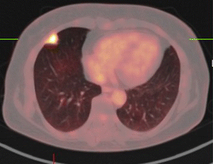
18F-FDG PET-CT combines functional (glucose metabolism) and anatomic (CT) imaging. It excels at staging lymphoma, lung, oesophageal, colorectal (restaging), cervical and head-and-neck cancers; assessing treatment response (Deauville score in lymphoma); and finding recurrence. Radiation dose is ~10–15 mSv.
FDG PET-CT injects 18F-fluorodeoxyglucose which accumulates in metabolically active cells — most cancers, but also inflammation and infection. Combined with contrast or non-contrast CT, it provides functional plus anatomical detail unmatched by either alone. Standard indications: lymphoma (staging and response — Deauville 1–5 scale), lung cancer (mediastinal staging + solitary pulmonary nodule > 8 mm), oesophageal cancer, cervical cancer, head-and-neck cancer, and colorectal cancer recurrence with rising CEA. Limitations: FDG is not tumour-specific — active infection, inflammation, brown fat, muscles and thymic rebound cause false positives. Some cancers are poorly FDG-avid (well-differentiated hepatocellular, prostate, mucinous, low-grade lymphomas). Total-body PET scanners are emerging that improve dose reduction and sensitivity. Preparation: fast 4–6 hours, restrict carbs 24 hours, warm room to minimise brown fat uptake, avoid strenuous exercise. Diabetes management is careful — hyperglycaemia reduces tumour uptake. Post-scan: hydrate, void frequently.
- Tracer
- 18F-FDG
- Dose
- ~10–15 mSv
- Best cancers
- Lymphoma, lung, oesophageal, cervical, colorectal
- Response scale
- Deauville 1–5 (lymphoma)
- False positives
- Infection, inflammation, brown fat
When PET-CT changes management
| Cancer | Common indication |
|---|---|
| Lymphoma | Staging + interim response + end-of-treatment |
| Lung | Solitary nodule > 8 mm; mediastinal staging |
| Oesophageal | Baseline staging + response |
| Cervical | Nodal + distant staging |
| Colorectal | Rising CEA / suspected recurrence |
Preparation
Fast 4–6 hours (water only).
Restrict carbohydrates 24 hours before to lower baseline muscle uptake.
Avoid strenuous exercise 24 hours before.
Blood glucose < 10 mmol/L needed (adjust insulin per specialist advice in diabetes).
Warm environment reduces brown-fat uptake — dress warmly.
- Incidental FDG-avid nodule outside known cancer — investigate for second primary.
- Diffuse thyroid uptake — thyroid cancer or thyroiditis workup.
- Colonic focal uptake in an asymptomatic patient — colonoscopy to exclude polyp/cancer.
PET-CT indication
- 1Suspected lymphoma?Staging PET-CT.
- 2Solitary pulmonary nodule ≥ 8 mm, indeterminate CT?PET-CT.
- 3Rising CEA post-colorectal treatment?PET-CT.
- 4Cervical cancer staging?PET-CT for nodal disease.
Related questions people ask
Frequently asked questions
- PET-CT combines function and anatomy.
- Excellent for lymphoma, lung, oesophageal and cervical staging.
- Deauville score standardises lymphoma response.
- Not tumour-specific — inflammation causes false positives.
- Preparation is critical for image quality.
References
2 sources- EANMFDG PET-CT Procedure Guidelines
European nuclear medicine.
eanm.org
- NCCN GuidelinesCancer-specific PET-CT indications
US oncology.
nccn.org
Related articles
 Imaging & Diagnostic Tests
Chest X-ray Explained: What It Shows, When You Need One, and Radiation Dose
A chest X-ray delivers a chest radiation dose equivalent to a few days of background radiation. Learn what it detects and what it misses.
 Imaging & Diagnostic Tests
CT Head Scan Explained: When It Is Needed for Stroke, Trauma and Headache
A non-contrast CT head is the first imaging test in stroke and head trauma. Learn NICE criteria, radiation dose, and what CT misses.
 Imaging & Diagnostic Tests
CTPA (CT Pulmonary Angiogram) Explained: Diagnosing Pulmonary Embolism
CTPA is the reference standard for pulmonary embolism. Learn indications, radiation dose, contrast risks, and pregnancy alternatives.