CT Abdomen and Pelvis Explained: Acute Abdomen, Contrast and Radiation Dose
CT abdomen/pelvis is the workhorse for acute abdominal pain. Learn contrast rules, radiation dose and when ultrasound is preferred.
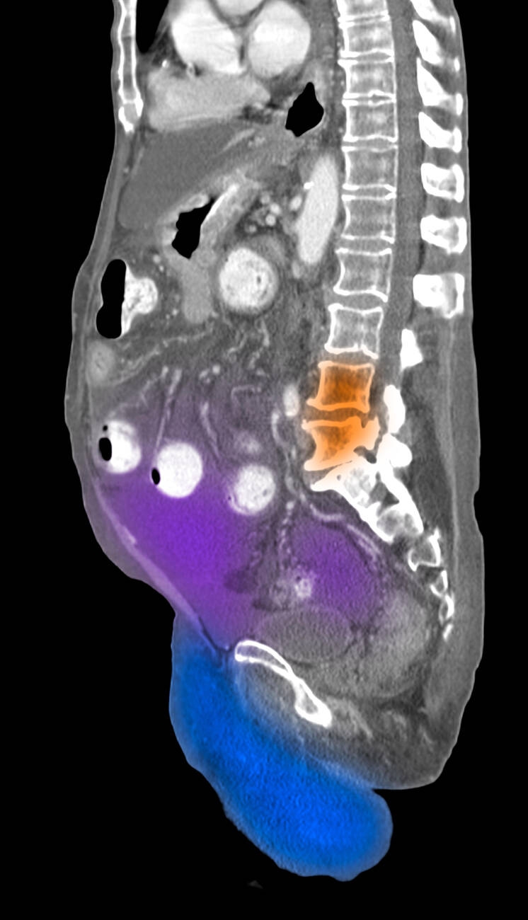
CT abdomen/pelvis with IV contrast is the standard for acute abdominal pain, staging cancer, and evaluating trauma. Radiation dose is ~8–10 mSv (~3 years background). Iodinated contrast is used, so check eGFR and allergy history. Ultrasound is preferred first-line in children, pregnancy, and for gallbladder/kidney assessment.
CT abdomen/pelvis has become the default cross-sectional test for acute abdominal complaints because of speed, breadth, and near-universal availability. Sensitivity for acute appendicitis > 95%, diverticulitis > 95%, bowel obstruction > 90%, mesenteric ischaemia (with arterial phase) 85–95%. Standard protocol uses IV iodinated contrast for optimum vascular and mural detail; oral contrast is largely obsolete in the acute setting except for suspected perforation or fistula. Consider unenhanced CT for renal colic (CT KUB — see linked article), acute haemorrhage on warfarin, and follow-up of known stones. ACR contrast-safety guidance permits IV contrast in eGFR ≥ 30 without pre-hydration; below 30, weigh benefit vs risk with a nephrologist. Metformin does not need routine holding unless AKI is expected. Post-contrast anaphylactoid reactions occur in ~0.6%; prior severe reaction merits premedication or alternative modality. Radiation dose can be halved with iterative reconstruction or dual-energy CT; children should be dosed on paediatric-adjusted protocols (Image Gently). Staging CTs follow tumour-specific NCCN protocols.
- Radiation dose
- ~8–10 mSv (~3 y background)
- Best for
- Acute abdomen, staging, trauma
- eGFR threshold
- ≥ 30 for contrast without pre-hydration
- Alternatives
- Ultrasound (kids, gallbladder), MRI (young/pregnant)
- Oral contrast
- Rarely needed in acute setting
CT vs ultrasound vs MRI for abdominal complaints
| Question | Best test |
|---|---|
| Right lower quadrant pain, adult | CT (or US in children/young women) |
| Right upper quadrant pain, gallstones | Ultrasound first |
| Suspected renal colic | CT KUB (low-dose) |
| Suspected Crohn/appendicitis in pregnancy | MRI abdomen |
| Cancer staging | CT + contrast (± PET-CT) |
Contrast safety
Check eGFR before contrast — reduce or omit if < 30 mL/min/1.73m².
Previous iodinated contrast reaction: premedicate with steroids + antihistamine or use MRI/US.
Diabetes on metformin: no routine hold if eGFR ≥ 30 and no AKI.
Pregnancy: IV iodinated contrast is generally avoided unless benefit outweighs risk.
- Acute abdominal pain + peritonism, sepsis, or shock — emergency CT.
- Suspected mesenteric ischaemia (out-of-proportion pain + acidosis) — CT angiogram.
- New anaemia + weight loss — CT staging pathway.
Acute abdominal pain workup
- 1Peritonism or sepsis?Urgent CT with contrast.
- 2Right upper quadrant + gallstones suspected?Start with ultrasound.
- 3Renal colic?Low-dose CT KUB (unenhanced).
- 4Suspected obstruction?CT (± oral contrast if fistula suspected).
Related questions people ask
Frequently asked questions
- CT abdomen/pelvis is the default for acute abdominal pain in adults.
- IV contrast is standard; check eGFR and allergy.
- Ultrasound and MRI are preferred in children and pregnancy.
- Oral contrast is rarely required now.
- Justify each scan — dose is real.
References
2 sources- ACR Manual on Contrast Media
US standard.
acr.org
- RCRStandards for intravascular contrast administration
UK standard.
rcr.ac.uk
Related articles
 Imaging & Diagnostic Tests
Chest X-ray Explained: What It Shows, When You Need One, and Radiation Dose
A chest X-ray delivers a chest radiation dose equivalent to a few days of background radiation. Learn what it detects and what it misses.
 Imaging & Diagnostic Tests
CT Head Scan Explained: When It Is Needed for Stroke, Trauma and Headache
A non-contrast CT head is the first imaging test in stroke and head trauma. Learn NICE criteria, radiation dose, and what CT misses.
 Imaging & Diagnostic Tests
CTPA (CT Pulmonary Angiogram) Explained: Diagnosing Pulmonary Embolism
CTPA is the reference standard for pulmonary embolism. Learn indications, radiation dose, contrast risks, and pregnancy alternatives.