Abdominal Ultrasound Explained: Uses, Preparation and Limitations
Ultrasound is safe, radiation-free, and excellent for gallstones, liver, kidneys and AAA. Learn its limits and when CT is needed.
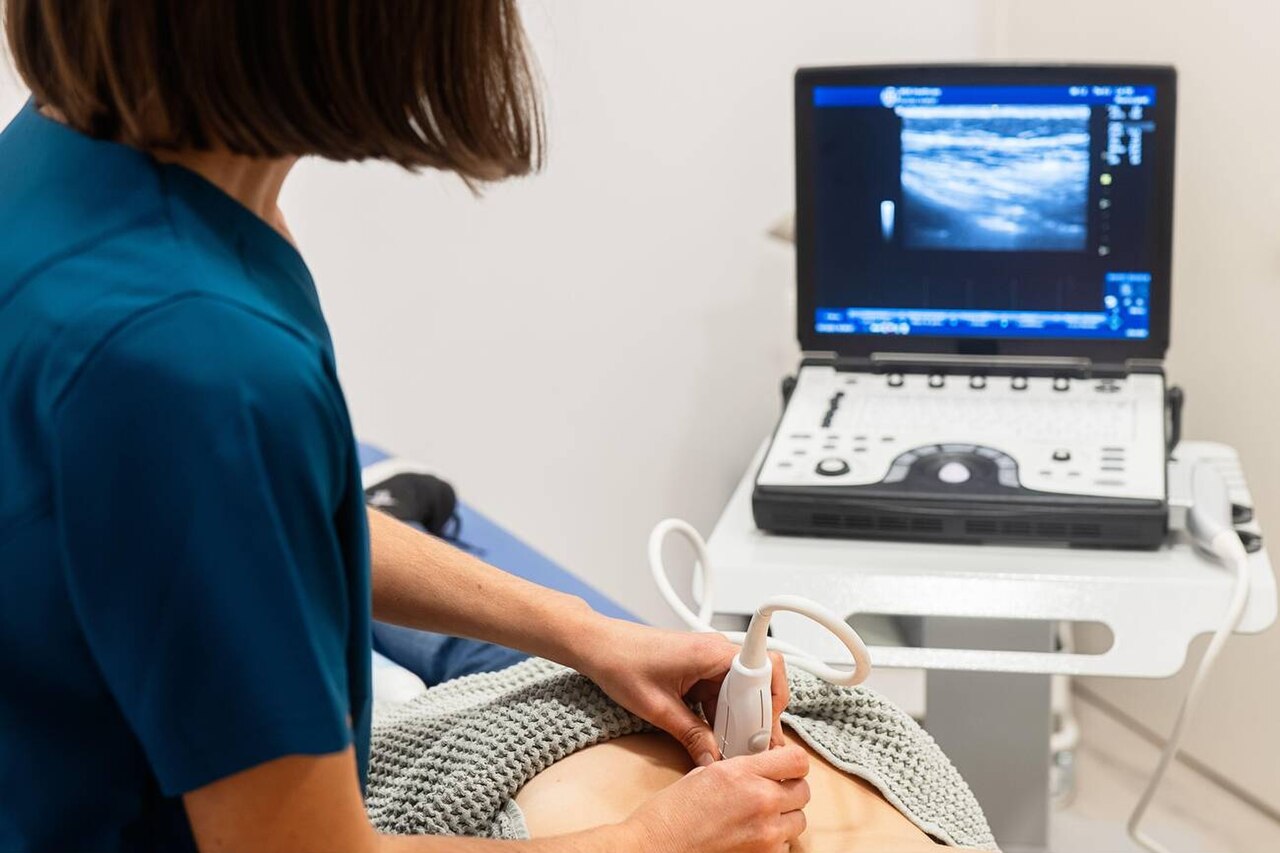
Abdominal ultrasound is safe, radiation-free, and excellent for gallstones, biliary dilatation, fatty liver, liver lesions, kidney stones/hydronephrosis, and abdominal aortic aneurysm (AAA). Preparation: fast 6 hours for gallbladder assessment; full bladder for pelvic views. Main limits: operator dependence, obesity, and bowel gas obscuring pancreas and bowel.
Ultrasound remains the first imaging test for most upper-abdominal complaints because it is fast, radiation-free, portable, and highly accurate for many structures. Gallstones: sensitivity > 95%, specificity > 95%. Fatty liver: sensitivity 60–94% depending on severity — mild steatosis (< 5%) may be missed. Hydronephrosis: sensitivity > 90% but ureteric stones themselves are frequently obscured; CT-KUB (low-dose) is now first-line for suspected ureteric colic. Aortic aneurysm screening (AAA): the UK NHS AAA Screening Programme invites men at 65; ultrasound has near-100% sensitivity for aneurysms > 3 cm. Limitations: dependent on operator experience; heavily obese patients (BMI > 40) reduce image quality; bowel gas frequently obscures pancreas — CT or MRI is preferred. Contrast-enhanced ultrasound (CEUS) can characterise liver lesions without radiation. Elastography (FibroScan, ARFI) quantifies liver stiffness for fibrosis assessment in NAFLD/MASLD.
- Radiation
- None
- Best for
- Gallstones, liver, kidneys, AAA
- Preparation
- Fast 6 h for GB; full bladder for pelvic
- Limits
- Obesity, bowel gas, operator dependence
- Ureteric stone?
- CT-KUB first-line (not USS)
When ultrasound vs alternative
| Question | Ultrasound | Alternative |
|---|---|---|
| Gallstones | First-line | MRCP if biliary obstruction |
| Abnormal LFTs | First-line | MRI/FibroScan for fibrosis |
| Ureteric stone | Limited | CT-KUB |
| Pancreatic lesion | Poor (gas) | CT/MRI |
| AAA screening | First-line | CT if repair being planned |
Typical findings and their meaning
Gallstones + wall thickening + pericholecystic fluid → acute cholecystitis.
Common bile duct > 6 mm (up to 8 mm post-cholecystectomy) → biliary obstruction.
Bright echogenic liver → hepatic steatosis (fatty liver).
Focal hyperechoic liver lesion → often haemangioma; characterise with CEUS or MRI if uncertain.
AAA — aortic diameter ≥ 3 cm; surveillance thresholds ≥ 4.5 cm every 3 months.
- Gallstones + fever + jaundice — cholangitis, urgent hospital assessment.
- AAA ≥ 5.5 cm — vascular referral for repair discussion.
- Hydronephrosis + fever — obstructed infected system, urgent decompression.
RUQ pain workup
- 1Colicky pain + gallstones on USS?Symptomatic cholelithiasis — cholecystectomy referral.
- 2Persistent fever + gallstones?Cholecystitis — admission, antibiotics.
- 3Jaundice + dilated CBD?MRCP + ERCP if stones.
- 4Raised ALT/AST + steatosis on USS?NAFLD/MASLD workup: FIB-4 + FibroScan.
Related questions people ask
Frequently asked questions
- Ultrasound is the first-line abdominal imaging for most complaints.
- Radiation-free and safe in pregnancy.
- Fast 6 h for gallbladder assessment.
- CT-KUB has replaced USS for ureteric stones.
- Obesity and bowel gas are the main limits.
References
2 sources- RCRiRefer
UK imaging referral guidance.
irefer.org.uk
- NHS AAA Screening Programme
UK national programme.
gov.uk
Related articles
 Imaging & Diagnostic Tests
Chest X-ray Explained: What It Shows, When You Need One, and Radiation Dose
A chest X-ray delivers a chest radiation dose equivalent to a few days of background radiation. Learn what it detects and what it misses.
 Imaging & Diagnostic Tests
CT Head Scan Explained: When It Is Needed for Stroke, Trauma and Headache
A non-contrast CT head is the first imaging test in stroke and head trauma. Learn NICE criteria, radiation dose, and what CT misses.
 Imaging & Diagnostic Tests
CTPA (CT Pulmonary Angiogram) Explained: Diagnosing Pulmonary Embolism
CTPA is the reference standard for pulmonary embolism. Learn indications, radiation dose, contrast risks, and pregnancy alternatives.