Mammogram Explained: Screening, BI-RADS, Dense Breasts and Recall
Screening mammography reduces breast-cancer mortality. Learn recall rates, BI-RADS categories, dense breasts, and when tomosynthesis or MRI is added.
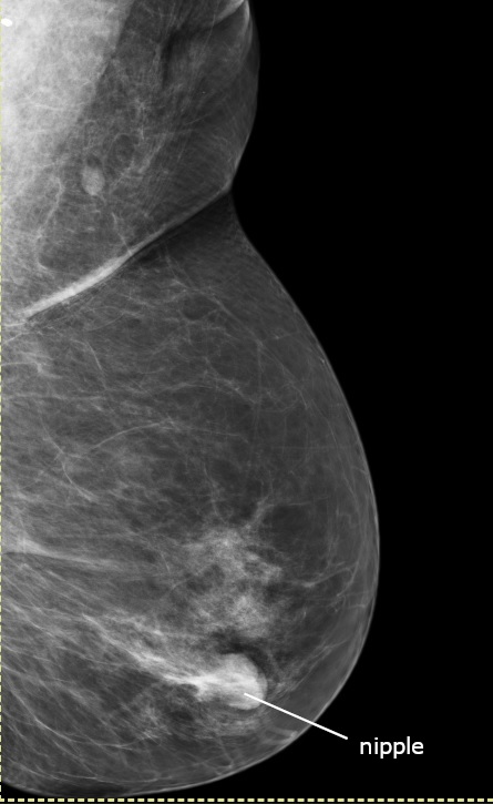
Screening mammography reduces breast-cancer mortality by ~20% in women 50–74. The NHS Breast Screening Programme invites women 50–71 every 3 years; the USPSTF updated 2024 recommendation is biennial from age 40. A recall does not mean cancer — most recalls (95%) are benign findings clarified by additional views or ultrasound.
Mammography uses low-dose X-rays (~0.4 mSv per bilateral view) to detect breast cancer at an earlier stage than clinical examination alone. UK: NHS invites women aged 50–70 (extending to 47–73 in the AgeX trial) every 3 years. US: USPSTF 2024 now recommends biennial screening from age 40; ACS suggests annual from 45 with option from 40. Digital breast tomosynthesis (3D mammography) modestly reduces recall and increases cancer detection compared with 2D digital mammography. Findings are reported using the ACR BI-RADS lexicon (0 incomplete → 6 known malignancy). Category 3 (probably benign) has < 2% malignancy and typically has short-term follow-up. Category 4/5 warrants biopsy. Dense breasts (BI-RADS density C or D) reduce mammographic sensitivity — supplemental screening ultrasound or MRI is offered in some services and states (US "dense-breast notification laws"). High-risk women (BRCA1/2 carriers, first-degree relative with breast cancer at young age, prior chest radiotherapy) are offered annual MRI + mammography starting age 25–30.
- Radiation dose
- ~0.4 mSv per bilateral study
- UK NHS interval
- Every 3 years, 50–71
- USPSTF (2024)
- Biennial from 40 (US)
- Recall rate
- ~4% (~95% benign)
- High-risk protocol
- MRI + mammogram from 25–30
BI-RADS categories
| Category | Meaning | Action |
|---|---|---|
| 0 | Incomplete | Additional imaging |
| 1 | Negative | Routine screening |
| 2 | Benign | Routine screening |
| 3 | Probably benign | 6-month follow-up (< 2% malignant) |
| 4 | Suspicious | Biopsy |
| 5 | Highly suggestive of malignancy | Biopsy |
| 6 | Known biopsy-proven malignancy | Treatment |
Dense breasts — what to know
Density is a mammographic finding, not a symptom.
Dense breast tissue can hide small cancers on 2D mammography.
Supplemental screening options: tomosynthesis (3D mammogram), whole-breast ultrasound, MRI for higher risk.
Discuss with your clinician if you have BI-RADS density C or D and additional risk factors.
- New breast lump, skin change, nipple discharge (bloody), or nipple retraction — same-week clinical assessment.
- BI-RADS 4/5 recall — biopsy pathway.
- Family history strongly suggestive of BRCA — genetics referral.
Screening decision
- 1Average risk?Follow national programme (UK 50–71 every 3 y; US biennial from 40).
- 2Dense breasts + family history?Discuss supplemental screening (US or MRI).
- 3BRCA1/2 or high-risk syndrome?Annual MRI + mammogram from 25–30.
Related questions people ask
Frequently asked questions
- Screening mammography reduces mortality in average-risk women.
- Recall does not equal cancer — 95% are benign.
- Dense breasts reduce sensitivity; supplemental options exist.
- BRCA and equivalent high-risk women need MRI + mammography from young age.
- Report BI-RADS 4/5 leads to biopsy.
References
3 sources- USPSTF 2024Breast Cancer Screening
US Preventive Services Task Force.
uspreventiveservicestaskforce.org
- NHS Breast Screening Programme
UK national programme.
gov.uk
- ACRBI-RADS Atlas
American College of Radiology.
acr.org
Related articles
 Imaging & Diagnostic Tests
Chest X-ray Explained: What It Shows, When You Need One, and Radiation Dose
A chest X-ray delivers a chest radiation dose equivalent to a few days of background radiation. Learn what it detects and what it misses.
 Imaging & Diagnostic Tests
CT Head Scan Explained: When It Is Needed for Stroke, Trauma and Headache
A non-contrast CT head is the first imaging test in stroke and head trauma. Learn NICE criteria, radiation dose, and what CT misses.
 Imaging & Diagnostic Tests
CTPA (CT Pulmonary Angiogram) Explained: Diagnosing Pulmonary Embolism
CTPA is the reference standard for pulmonary embolism. Learn indications, radiation dose, contrast risks, and pregnancy alternatives.