MRI Lumbar Spine Explained: Back Pain Red Flags and When Imaging Is Justified
Most back pain does not need imaging. Learn the NICE red flags that justify MRI and how to interpret incidental disc findings.
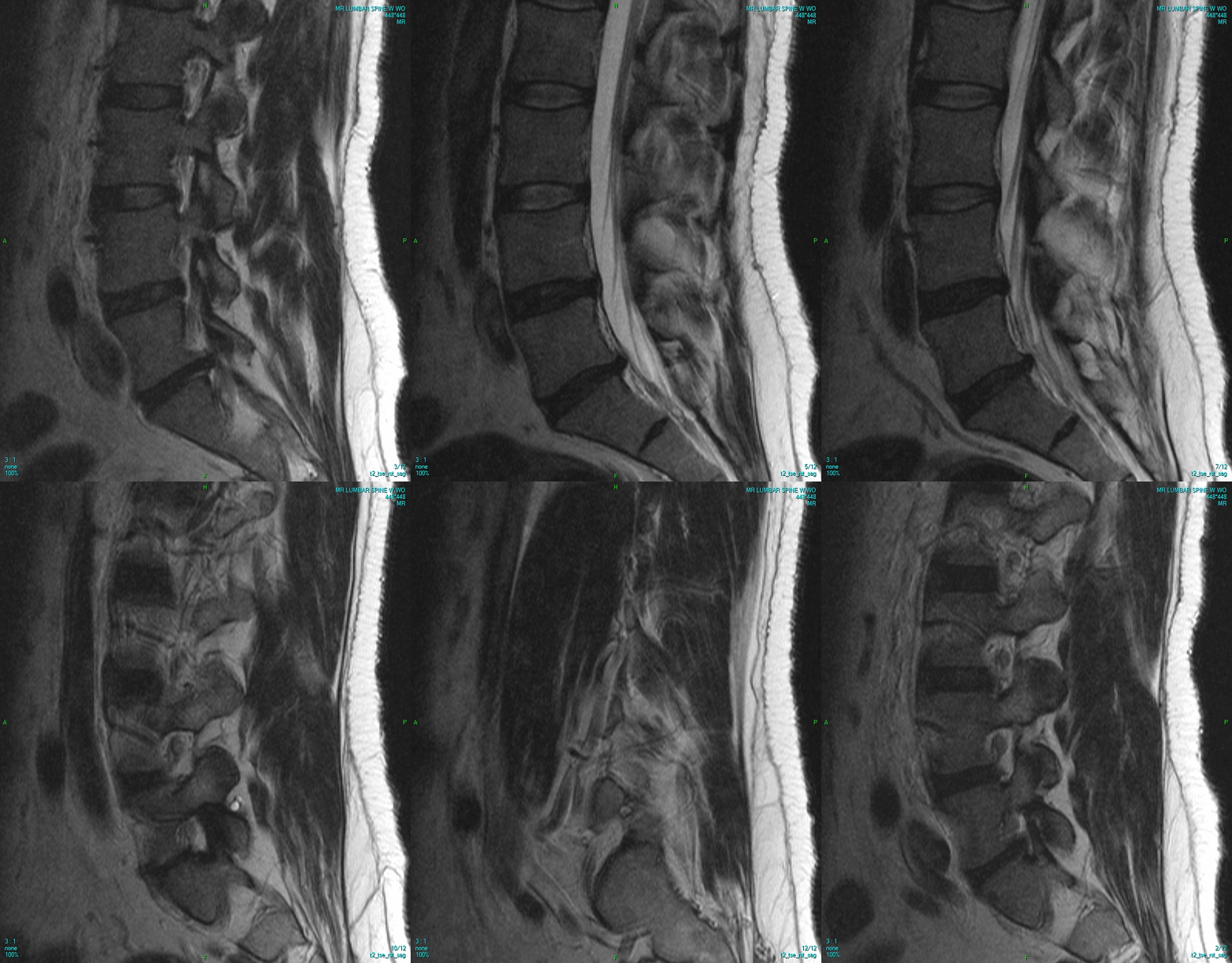
Most acute low back pain resolves without imaging. NICE and ACP recommend NO imaging in the first 6 weeks unless red flags: suspected cauda equina, cancer, infection, fracture, or progressive neurology. Routine MRI in uncomplicated back pain increases surgery and disability without benefit.
Lumbar spine MRI is highly sensitive but poorly specific — disc bulges, degeneration, and Modic changes are seen in 40–60% of asymptomatic adults over 40 and increase with age. NICE NG59 (2016, updated) and the American College of Physicians recommend AGAINST routine imaging for acute non-specific low back pain. Imaging is justified for red flags: suspected cauda equina (bilateral sciatica, saddle anaesthesia, bladder/bowel dysfunction — emergency MRI within hours); suspected malignancy (age > 50 with new pain, weight loss, night pain, history of cancer); suspected infection (fever, IVDU, immunosuppression, raised inflammatory markers); suspected fracture (major trauma, or minor trauma in osteoporosis/steroid use); progressive neurology; refractory sciatica > 6 weeks considering surgery. STIR sequences highlight oedema (infection/fracture); T2 shows nerve compression; contrast is added for suspected malignancy or infection. Reports commonly describe disc protrusions, Modic changes, facet arthropathy — normalise expectations: these findings are frequent in asymptomatic people.
- Radiation
- None
- Do NOT scan
- Uncomplicated back pain in first 6 weeks
- Scan when
- Red flags present
- Sensitivity for cauda equina
- Near 100%
- Asymptomatic disc bulge
- 40–60% of adults > 40 y
When MRI lumbar spine is justified
| Scenario | Imaging |
|---|---|
| Acute back pain < 6 weeks, no red flags | None — conservative management |
| Suspected cauda equina | Emergency MRI (< 4 h) |
| Cancer history + new back pain | MRI whole spine |
| Fever + back pain + raised CRP | MRI with contrast |
| Sciatica > 6 weeks, considering injection or surgery | MRI |
Red flags (NICE NG59)
Cauda equina symptoms: saddle anaesthesia, urinary retention/incontinence, faecal incontinence, bilateral sciatica.
Fever, IVDU, immunosuppression → spinal infection.
Age > 50 with new pain, weight loss, cancer history → malignancy.
Significant trauma or minor trauma in osteoporosis / steroid use → fracture.
Progressive weakness, foot drop, or loss of reflexes.
- Saddle numbness, new bladder/bowel dysfunction, or bilateral leg pain — same-day emergency assessment (cauda equina).
- Fever + back pain + raised CRP — hospital admission.
- Weight loss + night pain + history of cancer — urgent oncology imaging pathway.
Back pain imaging decision
- 1Any red flag?Urgent MRI.
- 2Radicular pain > 6 weeks despite treatment?MRI if considering procedure/surgery.
- 3Uncomplicated back pain?No imaging. Encourage activity + reassurance.
Related questions people ask
Frequently asked questions
- Do not image uncomplicated acute back pain in the first 6 weeks.
- Cauda equina needs same-day MRI.
- Age > 50 with new symptoms and cancer history triggers imaging.
- Disc bulges are common in asymptomatic people — beware overdiagnosis.
- STIR + contrast for infection or tumour workup.
References
2 sources- NICE NG59Low back pain and sciatica in over 16s
UK national guideline.
nice.org.uk
- ACPNoninvasive Treatments for Acute, Subacute, and Chronic Low Back Pain
American College of Physicians.
acpjournals.org
Related articles
 Imaging & Diagnostic Tests
Chest X-ray Explained: What It Shows, When You Need One, and Radiation Dose
A chest X-ray delivers a chest radiation dose equivalent to a few days of background radiation. Learn what it detects and what it misses.
 Imaging & Diagnostic Tests
CT Head Scan Explained: When It Is Needed for Stroke, Trauma and Headache
A non-contrast CT head is the first imaging test in stroke and head trauma. Learn NICE criteria, radiation dose, and what CT misses.
 Imaging & Diagnostic Tests
CTPA (CT Pulmonary Angiogram) Explained: Diagnosing Pulmonary Embolism
CTPA is the reference standard for pulmonary embolism. Learn indications, radiation dose, contrast risks, and pregnancy alternatives.