OCT Eye Scan Explained: Macula, Glaucoma and Diabetic Retinopathy
Optical coherence tomography images the retina in cross-section without contact. Learn its role in AMD, glaucoma and diabetic eye disease.
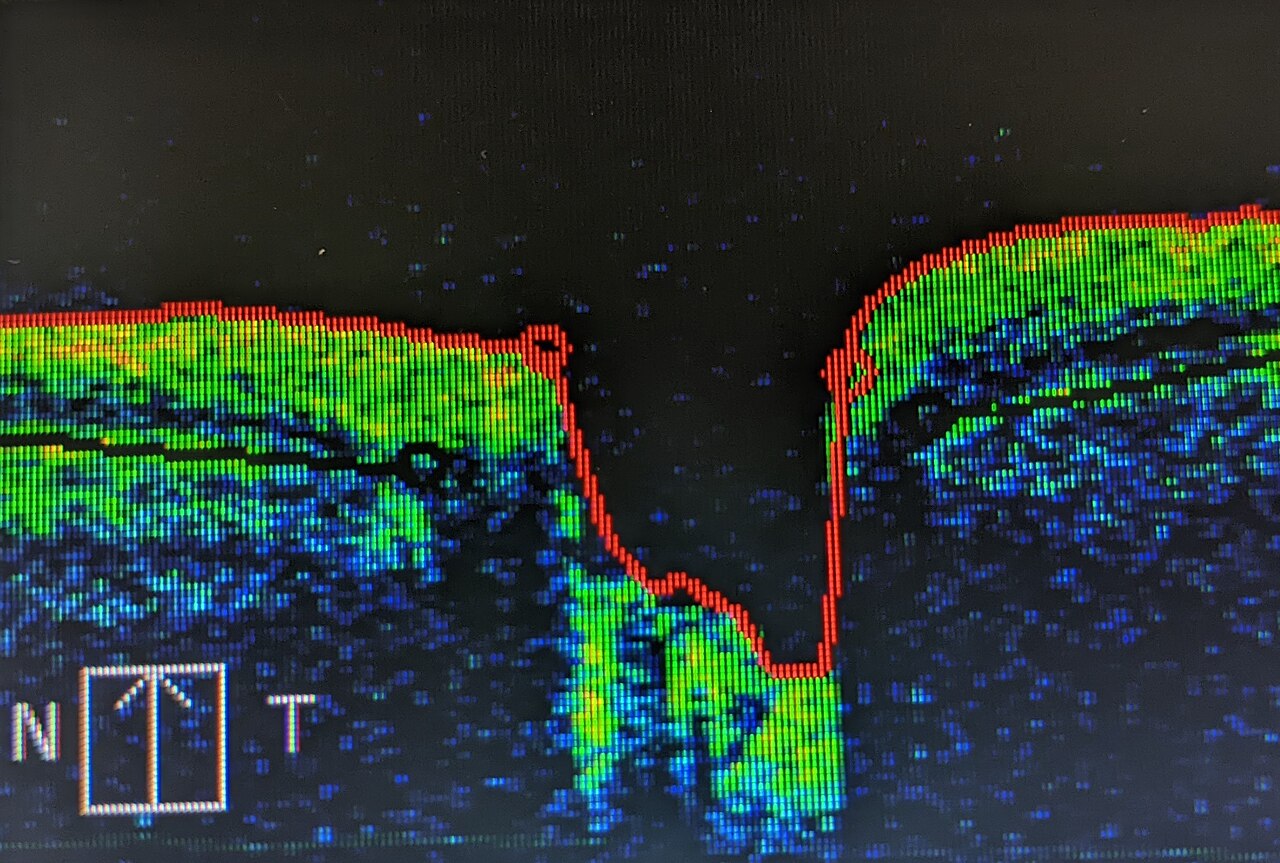
OCT (optical coherence tomography) is a rapid, non-contact scan producing high-resolution cross-sections of the retina. It is now standard for diagnosing and monitoring wet AMD, diabetic macular oedema, macular hole, epiretinal membrane, and glaucoma (retinal nerve fibre layer thickness). NHS diabetic eye screening uses OCT for referable maculopathy.
OCT has become the everyday backbone of retinal imaging. It uses low-coherence interferometry to build micron-resolution cross-sectional images of the retina and optic nerve head — non-contact, non-invasive, no radiation. Applications: (1) Age-related macular degeneration — detecting subretinal or intraretinal fluid triggers anti-VEGF injection therapy; treat-and-extend regimens rely on OCT. (2) Diabetic macular oedema — central retinal thickness > 300 µm triggers referral and treatment; NHS DES uses M1/M2 classifications. (3) Glaucoma — retinal nerve fibre layer (RNFL) and ganglion cell complex (GCC) thickness on Cirrus/Spectralis platforms track progression more sensitively than visual field. (4) Macular hole, epiretinal membrane, vitreomacular traction — surgical decisions. (5) Choroidal thickness — CSC and pachychoroid diseases. OCT angiography (OCT-A) adds vascular perfusion without dye, complementing fluorescein angiography in AMD and DR. Every optometrist practice in the UK now offers OCT; frequency 1–2 yearly for at-risk groups.
- Radiation
- None
- Contact
- None
- Key uses
- AMD, DMO, glaucoma, macular hole
- AMD marker
- Sub/intraretinal fluid
- DMO threshold
- Central retinal thickness > 300 µm
OCT in common conditions
| Condition | OCT finding |
|---|---|
| Wet AMD | Subretinal or intraretinal fluid |
| Diabetic macular oedema | Increased central retinal thickness |
| Glaucoma | Thinning of RNFL and GCC |
| Macular hole | Full-thickness break at the fovea |
| Epiretinal membrane | Thickened wrinkled internal limiting membrane |
Who benefits
Anyone with sudden central vision distortion or scotoma — same-day OCT to exclude wet AMD.
Diabetic patients — annual OCT via NHS DES if maculopathy detected on retinal photography.
Glaucoma suspects and confirmed patients — baseline + serial monitoring.
Post-cataract patients with unexplained visual disturbance — check for cystoid oedema.
- Sudden vision loss or central distortion — same-day ophthalmology.
- New macular fluid on OCT — anti-VEGF pathway.
- Central retinal thickness rising on OCT + diabetes — treatment pathway.
When OCT changes management
- 1Central vision distortion?OCT to exclude wet AMD.
- 2Diabetic maculopathy on photos?OCT to grade.
- 3Glaucoma suspect?RNFL/GCC OCT + visual field.
- 4Unexplained post-op vision?OCT for cystoid oedema.
Related questions people ask
Frequently asked questions
- OCT is non-contact, radiation-free, and standard in modern eye care.
- Detects AMD fluid, diabetic macular oedema, glaucoma thinning.
- Every high-street optometrist now offers OCT in the UK.
- Serial OCT drives anti-VEGF re-treatment.
- OCT-angiography extends to vessels.
References
2 sources- Royal College of OphthalmologistsClinical Guidelines
UK ophthalmology.
rcophth.ac.uk
- AAOPreferred Practice Patterns
American Academy of Ophthalmology.
aao.org
Related articles
 Imaging & Diagnostic Tests
Chest X-ray Explained: What It Shows, When You Need One, and Radiation Dose
A chest X-ray delivers a chest radiation dose equivalent to a few days of background radiation. Learn what it detects and what it misses.
 Imaging & Diagnostic Tests
CT Head Scan Explained: When It Is Needed for Stroke, Trauma and Headache
A non-contrast CT head is the first imaging test in stroke and head trauma. Learn NICE criteria, radiation dose, and what CT misses.
 Imaging & Diagnostic Tests
CTPA (CT Pulmonary Angiogram) Explained: Diagnosing Pulmonary Embolism
CTPA is the reference standard for pulmonary embolism. Learn indications, radiation dose, contrast risks, and pregnancy alternatives.