Audiometry Explained: Pure-Tone, Speech and Tympanometry
Audiometry maps your hearing across frequencies. Learn pure-tone thresholds, conductive vs sensorineural loss, and hearing-aid thresholds.
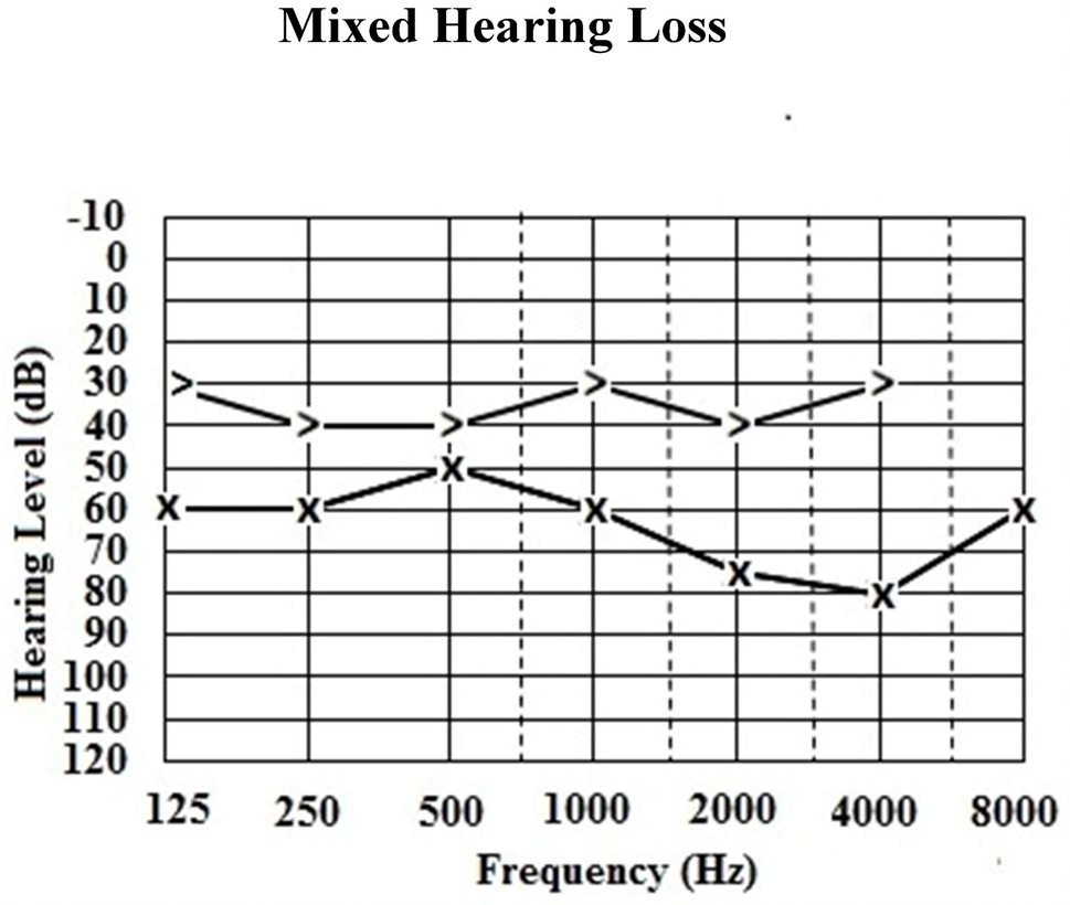
Pure-tone audiometry measures the softest sound you can hear at frequencies 250–8000 Hz through headphones (air conduction) and a bone vibrator (bone conduction). The gap between them separates conductive from sensorineural loss. Speech audiometry measures word discrimination. Tympanometry assesses middle-ear function.
Audiometry is the reference test for hearing loss classification. Pure-tone thresholds are plotted on an audiogram; normal ≤ 20 dB HL, mild 21–40 dB, moderate 41–70 dB, severe 71–90 dB, profound > 90 dB. Air–bone gap ≥ 10 dB → conductive component (otosclerosis, otitis media, cerumen, ossicular disruption). Sensorineural loss shows air = bone conduction thresholds. Asymmetric sensorineural loss (≥ 15 dB difference at any two adjacent frequencies) requires imaging (MRI internal auditory meatus) to exclude vestibular schwannoma. Speech audiometry: word recognition score < 60% suggests retrocochlear pathology. Tympanometry: type A normal, B flat (effusion/perforation), C negative pressure (Eustachian tube dysfunction). NICE NG98 (2018) recommends hearing assessment for any suspected hearing loss and 10-yearly for adults > 55 with risk factors. Otoacoustic emissions and auditory brainstem response are used in newborns and hard-to-test patients. Hearing-aid thresholds in NHS commonly ≥ 30 dB HL average.
- Normal hearing
- ≤ 20 dB HL
- Air–bone gap
- ≥ 10 dB = conductive
- Asymmetric SNHL
- MRI IAM to exclude schwannoma
- Tympanometry A
- Normal
- Tympanometry B
- Effusion or perforation
Types of hearing loss
| Pattern | Suggests |
|---|---|
| Symmetric high-frequency SNHL | Presbycusis |
| Cookie-bite mid-frequency SNHL | Congenital |
| Notch at 4 kHz | Noise-induced hearing loss |
| Conductive loss + type B tympanogram | Middle-ear effusion / perforation |
| Asymmetric SNHL | Vestibular schwannoma workup |
When to order audiometry
Any suspected hearing loss.
Tinnitus (single-sided especially — MRI thresholds apply).
Occupational noise exposure surveillance.
Sudden sensorineural hearing loss — emergency same-day audiogram + steroid pathway.
Balance disorders (asymmetric loss may indicate schwannoma).
- Sudden sensorineural hearing loss — audiogram + steroids within 3 days (best outcomes).
- Asymmetric hearing loss or unilateral tinnitus — MRI IAM.
- Persistent middle-ear effusion in adult — nasopharyngeal exam (exclude nasopharyngeal carcinoma).
Hearing loss workup
- 1Symmetric sensorineural age-related?Hearing aids if thresholds ≥ 30 dB average.
- 2Conductive loss + type B tympanogram?ENT for effusion / perforation.
- 3Asymmetric SNHL or single-sided tinnitus?MRI IAM.
- 4Sudden SNHL?Emergency steroid pathway.
Related questions people ask
Frequently asked questions
- Audiometry classifies hearing loss.
- Air–bone gap separates conductive from sensorineural.
- Asymmetric SNHL needs MRI IAM.
- Sudden SNHL is an emergency.
- NHS hearing-aid thresholds average ≥ 30 dB HL.
References
2 sources- NICE NG98Hearing loss in adults
UK national guideline.
nice.org.uk
- BSARecommended Procedures
British Society of Audiology.
thebsa.org.uk
Related articles
 Imaging & Diagnostic Tests
Chest X-ray Explained: What It Shows, When You Need One, and Radiation Dose
A chest X-ray delivers a chest radiation dose equivalent to a few days of background radiation. Learn what it detects and what it misses.
 Imaging & Diagnostic Tests
CT Head Scan Explained: When It Is Needed for Stroke, Trauma and Headache
A non-contrast CT head is the first imaging test in stroke and head trauma. Learn NICE criteria, radiation dose, and what CT misses.
 Imaging & Diagnostic Tests
CTPA (CT Pulmonary Angiogram) Explained: Diagnosing Pulmonary Embolism
CTPA is the reference standard for pulmonary embolism. Learn indications, radiation dose, contrast risks, and pregnancy alternatives.