EMG and Nerve Conduction Studies Explained: Neuropathy, Radiculopathy and Motor Neurone Disease
EMG and NCS locate the lesion in weakness and numbness. Learn how they distinguish neuropathy, radiculopathy, and MND.
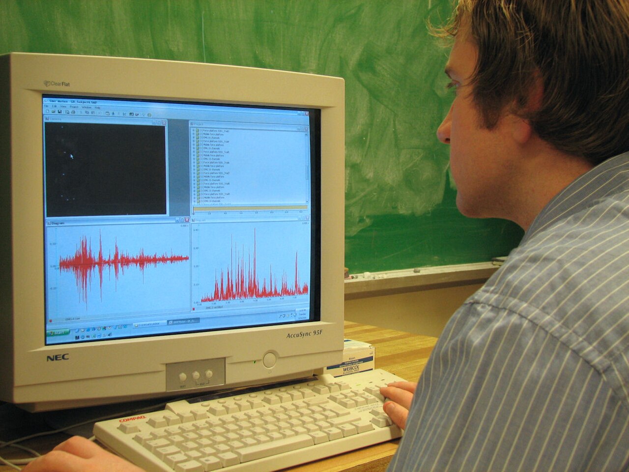
Nerve conduction studies (NCS) measure how well electrical impulses travel through peripheral nerves; electromyography (EMG) records the electrical activity of muscles. Together they localise weakness and numbness to muscle, neuromuscular junction, nerve root, plexus, or peripheral nerve, and distinguish axonal from demyelinating disease.
NCS and EMG are the reference bedside neurophysiology tests for peripheral neuromuscular symptoms. NCS: stimulates sensory or motor nerves and records amplitude, latency, and conduction velocity. Axonal neuropathy = low amplitude, relatively preserved conduction velocity. Demyelinating neuropathy (GBS, CIDP, hereditary demyelinating) = slowed conduction, prolonged distal latency, temporal dispersion, conduction block. EMG: needle electrode in muscle assesses spontaneous activity (fibrillations, positive sharp waves — signs of denervation) and motor unit potentials during volitional activation. Findings help localise: motor neurone disease shows widespread active denervation + reinnervation; radiculopathy shows changes limited to a myotome; carpal tunnel shows median sensory latency prolongation across the wrist. Common indications: suspected carpal tunnel, cubital tunnel, radiculopathy failing conservative management, diabetic neuropathy assessment, GBS/CIDP diagnosis, myopathy (with muscle biopsy), and motor neurone disease. Not necessary in classic clinical carpal tunnel with clear median distribution — treat empirically. Special: single-fibre EMG is the reference for myasthenia gravis; repetitive nerve stimulation confirms neuromuscular junction disorders.
- What NCS measures
- Amplitude, latency, conduction velocity
- What EMG measures
- Spontaneous + volitional muscle activity
- Axonal
- Low amplitude, preserved velocity
- Demyelinating
- Slowed velocity, conduction block
- MND
- Widespread denervation + reinnervation
NCS + EMG patterns
| Diagnosis | Typical findings |
|---|---|
| Carpal tunnel | Prolonged median distal sensory latency across wrist |
| Diabetic length-dependent neuropathy | Axonal, worst distal legs |
| CIDP | Multifocal demyelination with conduction block |
| Motor neurone disease | Widespread fibrillations + fasciculations + large motor units |
| L5 radiculopathy | Denervation in L5 myotome; normal sensory |
When to order
Progressive weakness, numbness, or paraesthesia after 4–6 weeks of symptoms.
Diagnosing carpal or cubital tunnel before surgery.
Suspected polyneuropathy — subtype and severity.
Suspected MND (fasciculations, atrophy, weakness).
Distinguishing radiculopathy from peripheral nerve entrapment.
- Rapidly progressive weakness — urgent neurology (Guillain-Barré pathway).
- Bulbar or respiratory symptoms + weakness — emergency admission.
- Sudden onset foot drop + back pain — cauda equina workup with MRI.
Weakness / numbness workup
- 1Focal + distribution suggests entrapment?NCS to localise.
- 2Distal symmetrical polyneuropathy?NCS + EMG to characterise axonal vs demyelinating.
- 3Suspected MND?Widespread NCS + EMG + neurology.
- 4Radiculopathy?MRI + EMG for functional confirmation.
Related questions people ask
Frequently asked questions
- NCS + EMG localise weakness and numbness.
- Axonal vs demyelinating distinction guides workup.
- Widespread denervation + reinnervation suggests MND.
- Carpal tunnel is the commonest referral.
- MRI complements EMG in radiculopathy.
References
2 sources- AANDistal Symmetric Polyneuropathy Practice Parameter
American Academy of Neurology.
aan.com
- AANEMPractice Guidelines
American Association of Neuromuscular & Electrodiagnostic Medicine.
aanem.org
Related articles
 Imaging & Diagnostic Tests
Chest X-ray Explained: What It Shows, When You Need One, and Radiation Dose
A chest X-ray delivers a chest radiation dose equivalent to a few days of background radiation. Learn what it detects and what it misses.
 Imaging & Diagnostic Tests
CT Head Scan Explained: When It Is Needed for Stroke, Trauma and Headache
A non-contrast CT head is the first imaging test in stroke and head trauma. Learn NICE criteria, radiation dose, and what CT misses.
 Imaging & Diagnostic Tests
CTPA (CT Pulmonary Angiogram) Explained: Diagnosing Pulmonary Embolism
CTPA is the reference standard for pulmonary embolism. Learn indications, radiation dose, contrast risks, and pregnancy alternatives.