Free T4 and Free T3: How to Read the Thyroid Panel Beyond TSH
TSH gets all the attention, but Free T4 and Free T3 are what actually tell you whether thyroid hormone is reaching your tissues. Learn what the patterns mean and when to test T3.
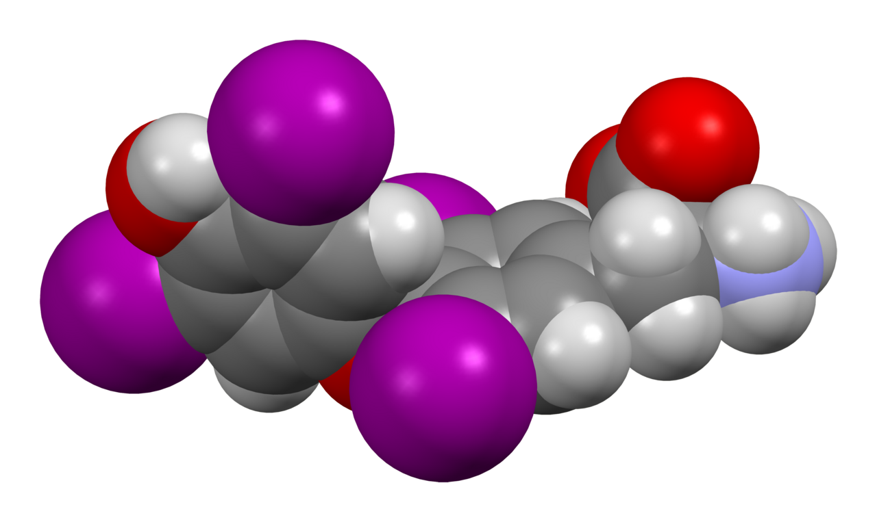
Free T4 (0.8–1.8 ng/dL or 12–22 pmol/L) is the storage hormone the thyroid releases. Free T3 (2.3–4.2 pg/mL or 3.5–6.5 pmol/L) is the active hormone tissues actually use. TSH tells you what the pituitary thinks; Free T4 and Free T3 tell you what your tissues are actually experiencing.
The thyroid releases mostly T4 (thyroxine); peripheral tissues convert it into the biologically active T3 (triiodothyronine). Measuring the free (unbound) fractions bypasses variation in binding proteins. Free T4 is the go-to confirmatory test after an abnormal TSH; Free T3 is added when hyperthyroidism is suspected (T3-toxicosis: TSH suppressed, Free T4 normal, Free T3 high) or when central hypothyroidism is being investigated. Non-thyroidal illness syndrome (sick euthyroid) — common in acute hospital patients — presents with low T3, sometimes low T4, and inappropriately normal or low TSH; do NOT diagnose primary thyroid disease during acute illness.
- Free T4 normal
- 0.8 – 1.8 ng/dL (12–22 pmol/L)
- Free T3 normal
- 2.3 – 4.2 pg/mL (3.5–6.5 pmol/L)
- When to add Free T3
- Suppressed TSH + normal Free T4 (T3 toxicosis)
- Reverse T3
- Rarely useful clinically — reflects non-thyroidal illness
- Do NOT interpret during
- Acute illness, hospital admission, or major surgery
What each hormone does
T4 (thyroxine) is 90% of the thyroid gland's output — but it is a storage form. It becomes biologically active only after peripheral deiodinases (5'-DIO1 and DIO2) convert it into T3 inside cells. This means T4 measurement tells you what the thyroid is releasing, while T3 measurement tells you what tissues are actually experiencing. Both hormones are 99% bound to plasma proteins (thyroxine-binding globulin, transthyretin, albumin) — measuring the free fraction is what makes the number physiologically interpretable.
The typical clinical workflow: order TSH first, add Free T4 if TSH is abnormal, add Free T3 if suspicion of hyperthyroidism is high with a normal Free T4, and consider TPO/Tg antibodies for autoimmune thyroid disease.
TSH + T4 + T3 pattern interpretation
| TSH | Free T4 | Free T3 | Diagnosis |
|---|---|---|---|
| High | Low | Low or normal | Overt primary hypothyroidism |
| High | Normal | Normal | Subclinical hypothyroidism |
| Low | High | High | Overt primary hyperthyroidism |
| Low | Normal | High | T3 toxicosis (early hyperthyroidism, T3-only) |
| Low | Normal | Normal | Subclinical hyperthyroidism |
| Low or normal | Low | Low | Central (pituitary) hypothyroidism OR non-thyroidal illness |
Why free measurements matter
Total T4 and total T3 include hormone bound to plasma proteins. Pregnancy raises thyroxine-binding globulin dramatically — total T4 doubles even though the free (active) fraction is stable. Oral oestrogen, oral contraceptives, and acute hepatitis do the same. Glucocorticoids, nephrotic syndrome, and severe illness lower binding proteins. Measuring the FREE fraction sidesteps all of this and is the modern standard.
Assay artefacts still exist: heterophile antibodies, biotin supplements, and dialysis membranes can produce misleading free hormone values. If a Free T4 result does not fit the clinical picture, ask the lab to repeat on a different assay platform.
When Free T3 is worth ordering
Free T3 is added in three specific situations: (1) TSH is suppressed and Free T4 is normal — screening for T3 toxicosis, which is common in Graves' disease and toxic nodules; (2) known hyperthyroidism is being monitored during treatment to catch relapse; (3) rare conditions such as thyroid hormone resistance or amiodarone-induced thyrotoxicosis. Routine ordering of Free T3 in every fatigue workup is not recommended — it adds cost and rarely changes management.
- Free T4 or Free T3 more than double the upper reference limit — thyroid storm risk if symptomatic.
- Free T4 very low with confusion, hypothermia, bradycardia — myxoedema coma risk.
- Suppressed TSH + new atrial fibrillation.
- Suppressed TSH + pregnancy (Graves' disease requires specialist care).
- Abnormal panel in a patient on amiodarone or lithium.
Interpreting a thyroid panel
- 1High TSH + low Free T4?Overt primary hypothyroidism — start levothyroxine and check TPO antibodies.
- 2High TSH + normal Free T4?Subclinical hypothyroidism. Repeat in 6–8 weeks. Treat if pregnant, symptomatic, or TPO-positive.
- 3Low TSH + high Free T4 or Free T3?Overt hyperthyroidism. Order TSH-receptor antibodies (Graves') and consider thyroid ultrasound or radioiodine uptake.
- 4Low TSH + normal Free T4 + normal Free T3?Subclinical hyperthyroidism. Repeat, screen for AF, review medications.
- 5Low TSH + low Free T4?Central (pituitary) hypothyroidism suspected — MRI pituitary, screen other axes.
- 6Panel drawn during acute illness?Non-thyroidal illness syndrome likely. Do NOT diagnose. Repeat 6–8 weeks after recovery.
Related questions people ask
- What is T3 toxicosis?
- When should Free T3 be ordered?
- How does TSH relate to Free T4?
- What is non-thyroidal illness syndrome?
- Do I need TPO antibodies?
- Can biotin affect Free T4?
- What is reverse T3?
Frequently asked questions
- TSH tells you what the pituitary is doing; Free T4 and Free T3 tell you what tissues experience.
- Free (not total) hormone is the modern standard — bypasses protein-binding artefacts.
- Add Free T3 when TSH is suppressed but Free T4 is normal (T3 toxicosis).
- Do NOT diagnose thyroid disease during acute illness — non-thyroidal illness syndrome mimics it.
- Reverse T3 is rarely useful clinically.
- Biotin supplements distort free hormone assays — pause 48 hours before the draw.
References
3 sources- ATAThyroid Function Testing Guidelines
American Thyroid Association testing recommendations.
thyroid.org
- NICE NG145Thyroid disease
UK thyroid diagnostic pathway.
nice.org.uk
- Endocrine SocietyThyroid Function During Pregnancy
Pregnancy-specific thyroid interpretation.
endocrine.org
Evidence Snapshot
This snapshot shows verified evidence records matched to this article. It is not a diagnosis or personal medical advice.
Thyroid peroxidase (TPO) antibodies are commonly found in autoimmune thyroid disease such as chronic autoimmune (Hashimoto's) thyroiditis and can also be present in a proportion of individuals with Graves' disease.
Limitation. A positive TPO antibody test can also occur in people without clinical thyroid disease, and prevalence varies by population.
TSH receptor antibodies (TRAb) are strongly associated with Graves' disease and are used, alongside clinical assessment and other tests, to help identify Graves' disease as the cause of hyperthyroidism.
Limitation. TRAb assay performance varies between laboratories, and antibody status alone does not describe disease activity or severity.
TSH reference ranges in pregnancy differ from those for non-pregnant adults because of the effects of human chorionic gonadotropin and other pregnancy-related changes, so trimester- and, where available, population-specific ranges are recommended.
Limitation. Ideal trimester-specific reference ranges vary between populations and assay methods, so a single universal range is not fully appropriate.
Related articles
 Laboratory Tests
TSH Test: Normal Range, High, Low & What Your Result Actually Means
TSH is the single most important thyroid number. Understand normal ranges by age and pregnancy, what a high or low value really tells you, and the two follow-up tests that clinch the diagnosis.
 Laboratory Tests
HbA1c Test: Normal Range, Prediabetic, Diabetic Cut-offs & How to Lower It
HbA1c reflects your average blood sugar over ~3 months. Understand every threshold — normal, prediabetes, diabetes — and the realistic actions that move the number.
 Laboratory Tests
LDL Cholesterol: Optimal Numbers, High Numbers, and How to Read Yours
LDL is the "bad" cholesterol only if you leave the story there. Understand optimal targets by risk group, the ApoB perspective, and the interventions that actually shift the number.