High Calcium with Normal PTH: The Non-Parathyroid Differential
A normal PTH does not rule out primary hyperparathyroidism — and it opens the door to malignancy, granulomatous disease, and vitamin D excess.
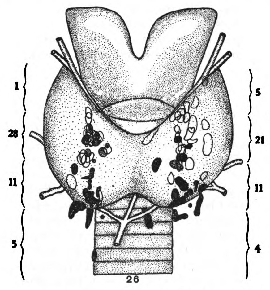
Hypercalcaemia with normal or low PTH points to a non-parathyroid cause — most commonly malignancy (PTHrP-mediated, bone metastases, or myeloma), granulomatous disease (sarcoidosis, TB), vitamin D excess, thiazide diuretics, or familial hypocalciuric hypercalcaemia. A normal PTH with high calcium is not normal — the PTH should be suppressed. The next step is urine calcium and a targeted differential.
Hypercalcaemia with normal or low PTH points to a non-parathyroid cause. Physiologically, high calcium should suppress PTH; a normal-range PTH is inappropriately normal and can still indicate mild primary hyperparathyroidism. Common non-PTH causes include malignancy, granulomatous disease, vitamin D excess, thiazide diuretics, and familial hypocalciuric hypercalcaemia.
- Expected PTH
- Suppressed when calcium high
- Inappropriately normal PTH
- Consider FHH or mild primary hyperparathyroidism
- Suppressed PTH
- Non-parathyroid cause — malignancy, granuloma, vit D
- Key next test
- 24-hour urine calcium + PTHrP
What normal PTH means when calcium is high
Physiologically, high calcium should drive PTH toward zero. A "normal-range" PTH in the presence of hypercalcaemia is therefore not truly normal — it is inappropriately normal, and can still indicate mild primary hyperparathyroidism. This is the most commonly missed diagnosis.
The urine calcium-to-creatinine clearance ratio distinguishes primary hyperparathyroidism (high urinary calcium) from familial hypocalciuric hypercalcaemia (low urinary calcium). The two look nearly identical on serum tests but require very different management.
PTH-independent hypercalcaemia — top causes
| Cause | Clues | Confirmatory test |
|---|---|---|
| Malignancy (PTHrP) | Rapid onset, weight loss, known cancer | PTHrP, imaging |
| Bone metastases / myeloma | Bone pain, anaemia, high protein | SPEP, imaging, calcium/creatinine ratio |
| Granulomatous disease | Sarcoidosis features, lung findings | ACE, chest imaging, 1,25(OH)D |
| Vitamin D toxicity | Supplement history, high 25(OH)D | 25-hydroxyvitamin D level |
| Thiazide diuretics | Recent medication | Trial off drug |
| Familial hypocalciuric hypercalcaemia | Family history, low urine Ca | CaSR genetic test |
| Milk-alkali syndrome | Calcium carbonate use | History |
- Calcium > 3.0 mmol/L (12 mg/dL) — hypercalcaemic crisis.
- New confusion, severe fatigue, or arrhythmia with high calcium.
- Rapid rise over weeks — think malignancy.
- Bone pain, unexplained fractures, anaemia — think myeloma.
Workup pathway
- 1PTH low, high calcium?PTH-independent — order PTHrP, SPEP, vitamin D (25 and 1,25), TSH, imaging.
- 2PTH normal or high, high calcium?Consider primary hyperparathyroidism vs FHH. Urine calcium-to-creatinine clearance ratio distinguishes.
- 3Very acute onset with known cancer?Hypercalcaemia of malignancy — urgent oncology involvement.
Related questions people ask
- What causes hypercalcaemia with low PTH?
- What is familial hypocalciuric hypercalcaemia?
- Can vitamin D toxicity cause high calcium?
- Do thiazide diuretics cause high calcium?
- What is PTHrP?
- When is a calcium level a medical emergency?
- Does dehydration raise calcium?
Frequently asked questions
- Normal PTH with high calcium is not normal — PTH should be suppressed.
- Non-PTH causes: malignancy, granuloma, vitamin D, thiazides, FHH.
- Urine calcium/creatinine ratio distinguishes primary HPT from FHH.
- Calcium > 3.0 mmol/L is an emergency.
References
2 sources- Endocrine Society GuidelinePrimary HPT
Diagnosis and management.
endocrine.org
- BMJ Best PracticeHypercalcaemia
Differential and workup.
bestpractice.bmj.com
Evidence Snapshot
This snapshot shows verified evidence records matched to this article. It is not a diagnosis or personal medical advice.
Total serum calcium is influenced by serum albumin, so a low albumin can lower total measured calcium without necessarily changing the biologically active ionised calcium fraction, which is why total calcium is often interpreted alongside albumin.
Limitation. Formulas for corrected calcium approximate but do not perfectly replace a direct ionised calcium measurement.
Symptoms of hypercalcaemia (such as fatigue, constipation, and confusion) and hypocalcaemia (such as muscle cramps and tingling) are nonspecific, so calcium abnormalities are often first detected on laboratory testing rather than from symptoms alone.
Limitation. Symptoms alone are neither sensitive nor specific for calcium abnormalities and cannot substitute for laboratory measurement.
Low serum magnesium can contribute to persistently low potassium and, in some cases, low calcium that do not correct as expected, because magnesium is required for normal handling of these other electrolytes.
Limitation. The clinical significance of borderline magnesium alterations varies by individual and situation.
Related articles
 Lab Interpretation
Vitamin D Deficiency: When Low Numbers Actually Matter
A "low" vitamin D result is not always a real deficiency, and not every low result needs treatment. Here is how clinicians actually decide.
 Lab Interpretation
High Ferritin with Low Iron: What the Combination Really Means
Ferritin is not just an iron store — it is also an inflammation marker. High ferritin with low serum iron usually means inflammation-trapped iron, not overload.
 Lab Interpretation
High HbA1c but Normal Blood Sugar Readings: What Is Going On?
Home glucose looks fine but the lab HbA1c is 6.5%? Post-meal spikes, dawn phenomenon, anaemia, and haemoglobin variants can all cause this paradox.