Thyroid Ultrasound Explained: TI-RADS, Nodules and When to Biopsy
Thyroid nodules are common and mostly benign. Learn ACR TI-RADS categories, biopsy thresholds, and how ultrasound guides management.
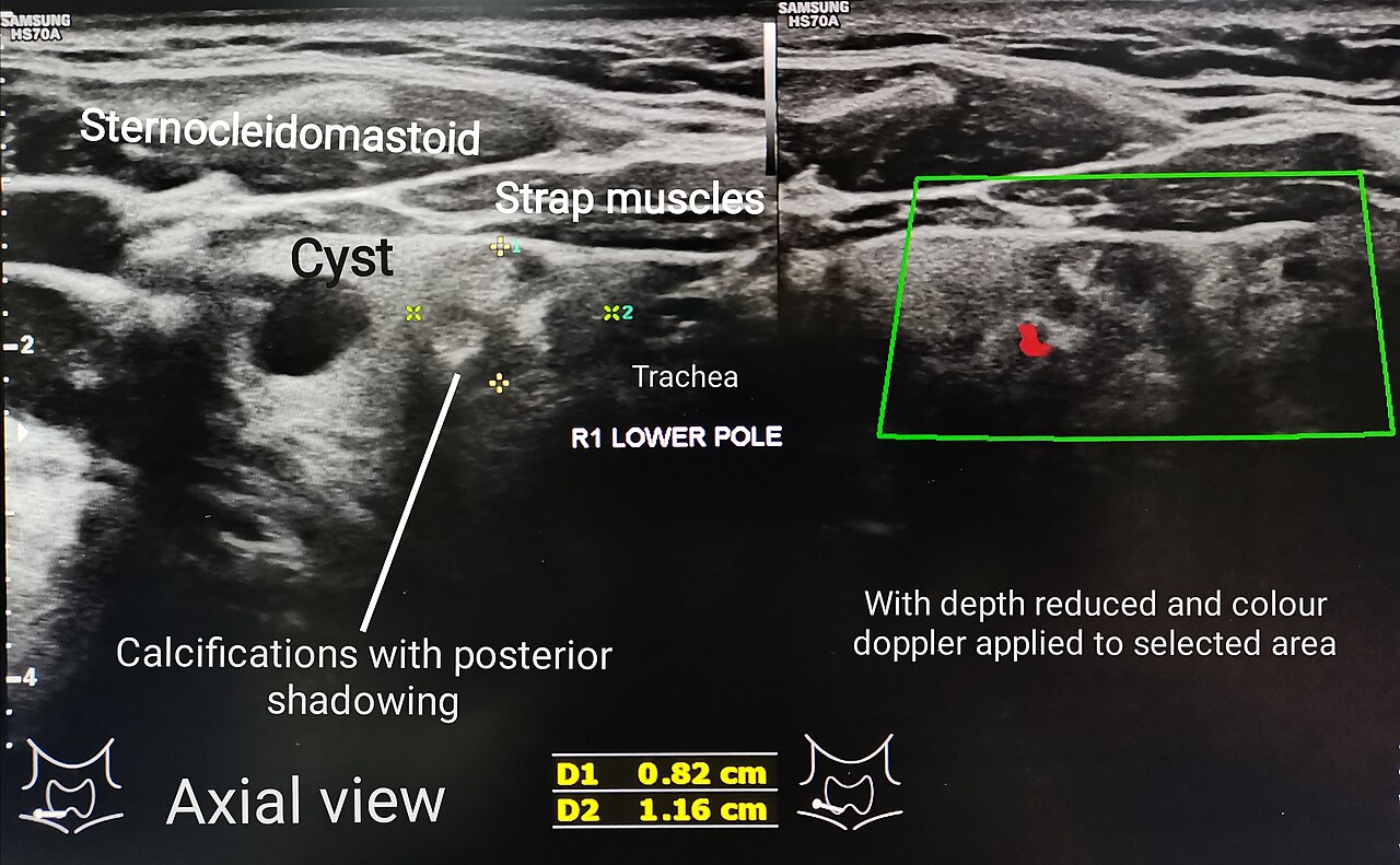
Thyroid nodules are found in up to 65% of adults on ultrasound but only 5–7% are malignant. ACR TI-RADS (Thyroid Imaging Reporting and Data System) scores nodules TR1 (benign) to TR5 (highly suspicious) based on composition, echogenicity, shape, margin, and echogenic foci. Biopsy is triggered by TI-RADS score AND size (e.g., TR5 ≥ 10 mm, TR4 ≥ 15 mm, TR3 ≥ 25 mm).
Thyroid ultrasound has become the primary imaging test for any palpable thyroid lump, thyroid dysfunction on biochemistry, or thyroid incidentaloma discovered on CT/MRI/PET. ACR TI-RADS (2017, updated 2023) uses a points system: five categories (composition, echogenicity, shape, margin, echogenic foci) score 0–3 points each, totalling TR1 (benign, 0 pts) to TR5 (highly suspicious, ≥ 7 pts). Size-based biopsy thresholds prevent over-biopsy: TR3 requires ≥ 25 mm; TR4 requires ≥ 15 mm; TR5 requires ≥ 10 mm (surveillance offered below these sizes). Fine-needle aspiration (FNA) is reported using the Bethesda system (I–VI). ATA 2015 recommends against biopsy of purely cystic nodules and any nodule < 10 mm without high-risk features. TSH must always be checked alongside — hyperfunctioning (hot) nodules on scintigraphy are almost never malignant and typically do not need FNA. Elastography (thyroid stiffness) is emerging as an adjunct. Molecular testing (Afirma, ThyroSeq) is used for Bethesda III/IV indeterminate FNAs to reduce diagnostic lobectomies.
- Radiation
- None
- Reporting
- ACR TI-RADS TR1–TR5
- Preparation
- None
- Adjunct
- TSH + thyroid antibodies + calcitonin (if MTC)
- FNA reporting
- Bethesda I–VI
TI-RADS categories + biopsy thresholds
| Category | Suspicion | Biopsy at |
|---|---|---|
| TR1 | Benign | No FNA |
| TR2 | Not suspicious | No FNA |
| TR3 | Mildly suspicious | ≥ 25 mm (surveillance ≥ 15 mm) |
| TR4 | Moderately suspicious | ≥ 15 mm (surveillance ≥ 10 mm) |
| TR5 | Highly suspicious | ≥ 10 mm (surveillance ≥ 5 mm) |
When ultrasound is needed
Palpable thyroid nodule or lump.
Thyroid dysfunction on biochemistry (subacute thyroiditis assessment).
Incidentaloma on CT/MRI/PET (any nodule > 10 mm merits US).
Follow-up of known nodules per TI-RADS surveillance schedule.
Preoperative mapping and lymph-node staging in known thyroid cancer.
- Rapidly growing thyroid lump — urgent 2-week referral.
- Hoarseness, dysphagia, or fixation of nodule — urgent ENT/endocrine.
- Thyroid nodule + suspicious cervical nodes — full-neck US + FNA.
Thyroid nodule workup
- 1Palpable nodule or incidentaloma > 10 mm?Ultrasound + TSH.
- 2Hyperfunctioning (low TSH)?Scintigraphy; hot nodules seldom malignant.
- 3TI-RADS + size meets biopsy criteria?FNA under US guidance (Bethesda).
- 4Bethesda III/IV?Consider molecular testing before surgery.
Related questions people ask
Frequently asked questions
- Thyroid ultrasound is first-line for any thyroid nodule.
- ACR TI-RADS standardises risk assessment.
- Biopsy thresholds prevent over-investigation.
- Always check TSH alongside.
- 95% of nodules are benign.
References
2 sources- ACR TI-RADS 2017 (updated 2023)
US reporting system.
acr.org
- ATA 2015 Thyroid Nodules and Cancer Guidelines
US thyroid association.
liebertpub.com
Related articles
 Imaging & Diagnostic Tests
Chest X-ray Explained: What It Shows, When You Need One, and Radiation Dose
A chest X-ray delivers a chest radiation dose equivalent to a few days of background radiation. Learn what it detects and what it misses.
 Imaging & Diagnostic Tests
CT Head Scan Explained: When It Is Needed for Stroke, Trauma and Headache
A non-contrast CT head is the first imaging test in stroke and head trauma. Learn NICE criteria, radiation dose, and what CT misses.
 Imaging & Diagnostic Tests
CTPA (CT Pulmonary Angiogram) Explained: Diagnosing Pulmonary Embolism
CTPA is the reference standard for pulmonary embolism. Learn indications, radiation dose, contrast risks, and pregnancy alternatives.