Venous Doppler Ultrasound Explained: Diagnosing DVT of the Leg
Compression ultrasound is the reference standard for DVT. Learn Wells DVT gating, above- vs below-knee scanning and repeat protocols.
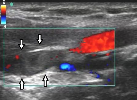
Compression ultrasound (venous Doppler) is the reference standard for suspected DVT. Sensitivity for proximal (above-knee) DVT is > 95%. Isolated distal (below-knee) DVT is less reliably imaged — some services scan whole-leg; others do proximal-only and repeat in 5–7 days if D-dimer positive and initial scan negative.
Venous Doppler ultrasound uses B-mode compressibility (a normal vein compresses under probe pressure; a clotted vein does not) plus colour and pulsed-wave Doppler. NICE NG158 and ISTH recommend: two-tier Wells DVT score gates D-dimer and imaging. Wells ≤ 1 (unlikely) + negative D-dimer → DVT excluded. Wells ≥ 2 (likely) or D-dimer positive → proximal compression ultrasound. Two approaches to below-knee DVT: (1) whole-leg ultrasound to detect isolated calf DVT; (2) proximal-only ultrasound with a repeat in 5–7 days if D-dimer positive — outcomes are equivalent. Whole-leg scanning is more sensitive but yields more false positives (small calf clots often self-resolve). Chronic post-thrombotic changes (residual venous obstruction, valvular reflux) are important in recurrent DVT workup. Ultrasound cannot distinguish acute from chronic DVT reliably — clinical history + comparison with prior scans matters. Upper-limb DVT (Paget–Schroetter, catheter-related) uses similar principles. IVC/pelvic vein thrombosis requires CT or MR venography.
- Radiation
- None
- Sensitivity proximal DVT
- > 95%
- Wells score gates
- D-dimer and imaging
- Whole-leg vs proximal
- Both accepted (repeat if proximal)
- Chronic vs acute
- Compare with prior scans
DVT diagnostic pathways
| Wells score | D-dimer | Next step |
|---|---|---|
| Unlikely (≤ 1) | Negative | DVT excluded |
| Unlikely (≤ 1) | Positive | Compression ultrasound |
| Likely (≥ 2) | N/A | Compression ultrasound within 4 h (or D-dimer + repeat) |
| Proximal US negative + high suspicion | N/A | Repeat US in 5–7 days |
Common findings + pitfalls
Non-compressible venous segment = acute DVT.
Echogenic thickened wall with partial compression = chronic post-thrombotic change.
Baker's cyst rupture is a classic mimic — visualise the cyst.
Superficial thrombophlebitis often coexists but is a different entity.
- Painful swollen leg + suspected DVT — compression US within 4 h (NICE NG158).
- DVT + breathlessness or pleuritic pain — PE workup.
- Iliofemoral DVT + severe symptoms — vascular MDT for catheter-directed thrombolysis.
Suspected DVT workup
- 1Wells score?Unlikely + neg D-dimer → excluded. Otherwise proximal US.
- 2Proximal US positive?Anticoagulation.
- 3Proximal US negative + still concerned?Repeat US in 5–7 days.
- 4Suspected iliofemoral or IVC extension?CT/MR venography.
Related questions people ask
Frequently asked questions
- Compression US is the reference standard for DVT.
- Wells score gates D-dimer and imaging.
- Proximal-only with repeat is a valid alternative to whole-leg.
- Ultrasound is safe in pregnancy.
- Iliofemoral extension needs CT/MR venography.
References
2 sources- NICE NG158VTE Diseases
UK national guideline.
nice.org.uk
- ACR Appropriateness CriteriaSuspected DVT
US imaging appropriateness.
acr.org
Related articles
 Imaging & Diagnostic Tests
Chest X-ray Explained: What It Shows, When You Need One, and Radiation Dose
A chest X-ray delivers a chest radiation dose equivalent to a few days of background radiation. Learn what it detects and what it misses.
 Imaging & Diagnostic Tests
CT Head Scan Explained: When It Is Needed for Stroke, Trauma and Headache
A non-contrast CT head is the first imaging test in stroke and head trauma. Learn NICE criteria, radiation dose, and what CT misses.
 Imaging & Diagnostic Tests
CTPA (CT Pulmonary Angiogram) Explained: Diagnosing Pulmonary Embolism
CTPA is the reference standard for pulmonary embolism. Learn indications, radiation dose, contrast risks, and pregnancy alternatives.