Visual Field Test (Perimetry) Explained: Glaucoma, Stroke and Chiasmal Lesions
Perimetry maps peripheral vision. Learn what patterns suggest glaucoma, stroke, or chiasmal lesions, and interpret Humphrey printouts.
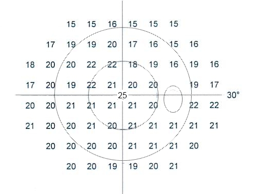
Perimetry (automated visual field test) maps how well you see across the visual field. It is essential for glaucoma diagnosis and monitoring and for detecting neurological visual field defects (chiasmal, retrochiasmal lesions). Humphrey printouts report MD, PSD, VFI and GHT — key values to check.
Automated static perimetry (Humphrey Field Analyzer, Octopus) is a psychophysical test requiring reliable patient responses to light stimuli of varying intensity across the visual field. Common protocols: 24-2 for glaucoma screening/monitoring, 30-2 for wider testing, 10-2 for advanced glaucoma or macular pathology. Key indices: Mean Deviation (MD, overall sensitivity vs age-matched normal), Pattern Standard Deviation (PSD, focal defects), Visual Field Index (VFI, 0–100% for progression tracking), Glaucoma Hemifield Test (GHT, "outside normal limits" flags). Reliability indices: fixation losses (< 20%), false positives (< 15%), false negatives (< 15%). Patterns: glaucoma — arcuate scotomas, nasal step, paracentral defect; superior altitudinal — anterior ischaemic optic neuropathy; central scotoma — optic neuritis, toxic/nutritional; bitemporal hemianopia — chiasmal (pituitary) lesion; homonymous hemianopia — retrochiasmal (stroke, tumour). Perimetry drives glaucoma treatment escalation — Guided Progression Analysis (GPA) confirms progression. NICE NG81 (2017, updated) recommends perimetry at glaucoma diagnosis and follow-up (usually 6–12 monthly).
- Common protocol
- 24-2 (glaucoma)
- MD
- Overall sensitivity deviation
- PSD
- Focal defect measure
- GHT
- "Outside normal limits" flag
- Reliability
- Fixation loss < 20%
Field defect patterns
| Pattern | Suggests |
|---|---|
| Arcuate scotoma respecting horizontal midline | Glaucoma |
| Superior altitudinal | AION |
| Central scotoma | Optic neuritis, toxic/nutritional |
| Bitemporal hemianopia | Chiasmal lesion (pituitary) |
| Homonymous hemianopia | Retrochiasmal (stroke, tumour) |
When to order
Glaucoma diagnosis and monitoring (baseline + 6–12 monthly).
Suspected chiasmal or retrochiasmal lesion.
Optic neuritis or optic neuropathy.
Retinal detachment or macular disease.
Screening for hydroxychloroquine retinopathy (10-2).
- Sudden new field defect — urgent neuroimaging.
- Progressive glaucoma field loss on GPA — treatment escalation.
- Bitemporal hemianopia — pituitary MRI.
When perimetry changes management
- 1Glaucoma?Baseline + 6–12 monthly.
- 2Chiasmal pattern?MRI pituitary.
- 3Homonymous defect?Neuro-imaging.
- 4Hydroxychloroquine user > 5 y?10-2 annual screening.
Related questions people ask
Frequently asked questions
- Perimetry maps peripheral vision.
- Essential in glaucoma diagnosis and monitoring.
- Field patterns can localise CNS lesions.
- Bitemporal hemianopia = chiasmal until proven otherwise.
- Reliability indices matter for interpretation.
References
2 sources- NICE NG81Glaucoma: diagnosis and management
UK national guideline.
nice.org.uk
- EGSEuropean Glaucoma Society Terminology and Guidelines
European glaucoma society.
eugs.org
Related articles
 Imaging & Diagnostic Tests
Chest X-ray Explained: What It Shows, When You Need One, and Radiation Dose
A chest X-ray delivers a chest radiation dose equivalent to a few days of background radiation. Learn what it detects and what it misses.
 Imaging & Diagnostic Tests
CT Head Scan Explained: When It Is Needed for Stroke, Trauma and Headache
A non-contrast CT head is the first imaging test in stroke and head trauma. Learn NICE criteria, radiation dose, and what CT misses.
 Imaging & Diagnostic Tests
CTPA (CT Pulmonary Angiogram) Explained: Diagnosing Pulmonary Embolism
CTPA is the reference standard for pulmonary embolism. Learn indications, radiation dose, contrast risks, and pregnancy alternatives.