Ambulatory ECG Explained: Holter, Event Recorder and Implantable Loop Recorder
Not every arrhythmia shows on a standard ECG. Learn Holter monitors, patch ECGs, event recorders and implantable loop recorders.
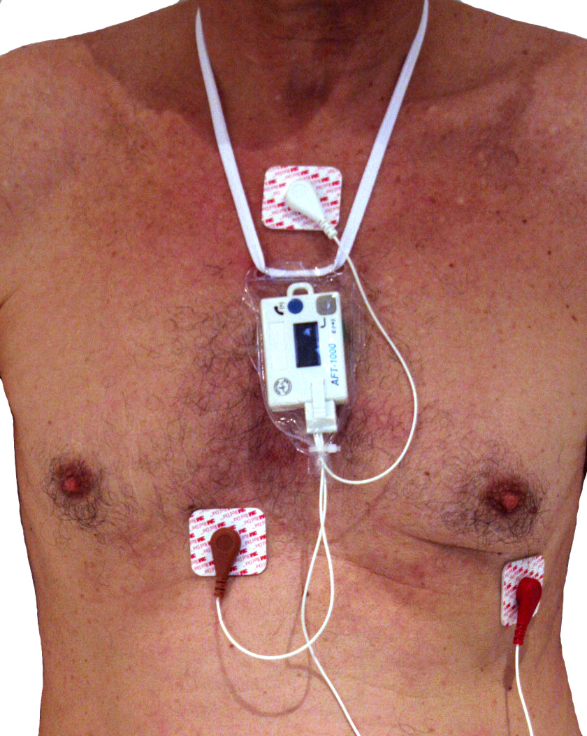
A single 12-lead ECG catches only current rhythms. Ambulatory ECG (Holter 24–72 h, patch 7–30 days, event recorder, or implantable loop recorder for weeks to years) extends detection for palpitations, unexplained syncope, and post-cryptogenic-stroke AF screening. Choice depends on symptom frequency.
Ambulatory ECG monitoring extends the detection window when arrhythmias are paroxysmal. Options: (1) Holter monitor — continuous 24–72 h; best for daily symptoms. (2) Patch ECG (Zio, Bardy) — continuous 7–14 (up to 30) days; best for weekly symptoms and post-stroke AF screening. (3) External event recorder — patient-triggered or auto-triggered over 30 days; best for infrequent but reproducible palpitations. (4) Implantable loop recorder (ILR) — subcutaneous device recording for up to 3 years; best for infrequent syncope (ESC 2018 syncope guideline endorses ILR early after unexplained syncope). ILR increased detection of AF after cryptogenic stroke from 1.4% to 12.4% in CRYSTAL-AF (NEJM 2014) and now supports secondary prevention with anticoagulation. NICE recommends prolonged monitoring (≥ 72 h or patch) after cryptogenic stroke to guide antithrombotic decisions. Symptom-diary correlation is crucial: patients log activities that align with recorded events.
- Holter
- 24–72 h continuous
- Patch
- 7–30 days continuous
- Event recorder
- 30 days patient-triggered
- ILR
- Up to 3 years subcutaneous
- Cryptogenic stroke
- Prolonged monitoring per NICE/ESC
Which ambulatory ECG for which scenario
| Scenario | Best test |
|---|---|
| Daily palpitations | 24–48 h Holter |
| Weekly palpitations | 7–14 day patch |
| Monthly episodes | Event recorder |
| Rare syncope | ILR |
| Cryptogenic stroke workup | Prolonged monitoring or ILR |
Symptom-recording tips
Keep a diary — record symptom onset, duration, and activity when they occur.
Do not stop your usual activities — the monitor is designed to be worn normally.
Avoid MRI, magnets, and immersion in water unless device is waterproof.
Report severe or persistent symptoms — do not wait until scan review.
- Syncope with injury or during exertion — urgent cardiology.
- Recurrent unexplained syncope — ILR consideration.
- Newly diagnosed AF — anticoagulation and rate/rhythm control planning.
Palpitation / syncope workup
- 1Palpitations recurrent daily?24–72 h Holter.
- 2Episodes weekly?7–14 day patch.
- 3Unexplained syncope + high recurrence risk?ILR.
- 4Post-cryptogenic stroke?Prolonged monitor or ILR to detect AF.
Related questions people ask
Frequently asked questions
- Match monitor duration to symptom frequency.
- ILR detects rare but important arrhythmias.
- Diary correlation matters.
- Prolonged monitoring guides post-stroke anticoagulation.
- Smartwatches augment but do not replace clinical monitoring.
References
2 sources- ESC 2018 Syncope Guideline
European cardiology.
academic.oup.com
- NICEAtrial fibrillation: diagnosis and management (NG196)
UK national guideline.
nice.org.uk
Related articles
 Imaging & Diagnostic Tests
Chest X-ray Explained: What It Shows, When You Need One, and Radiation Dose
A chest X-ray delivers a chest radiation dose equivalent to a few days of background radiation. Learn what it detects and what it misses.
 Imaging & Diagnostic Tests
CT Head Scan Explained: When It Is Needed for Stroke, Trauma and Headache
A non-contrast CT head is the first imaging test in stroke and head trauma. Learn NICE criteria, radiation dose, and what CT misses.
 Imaging & Diagnostic Tests
CTPA (CT Pulmonary Angiogram) Explained: Diagnosing Pulmonary Embolism
CTPA is the reference standard for pulmonary embolism. Learn indications, radiation dose, contrast risks, and pregnancy alternatives.