CT KUB Scan Explained: Diagnosing Kidney Stones and Ureteric Colic
CT KUB is the reference standard for renal colic. Learn NICE thresholds, radiation dose, and when ultrasound is preferred.
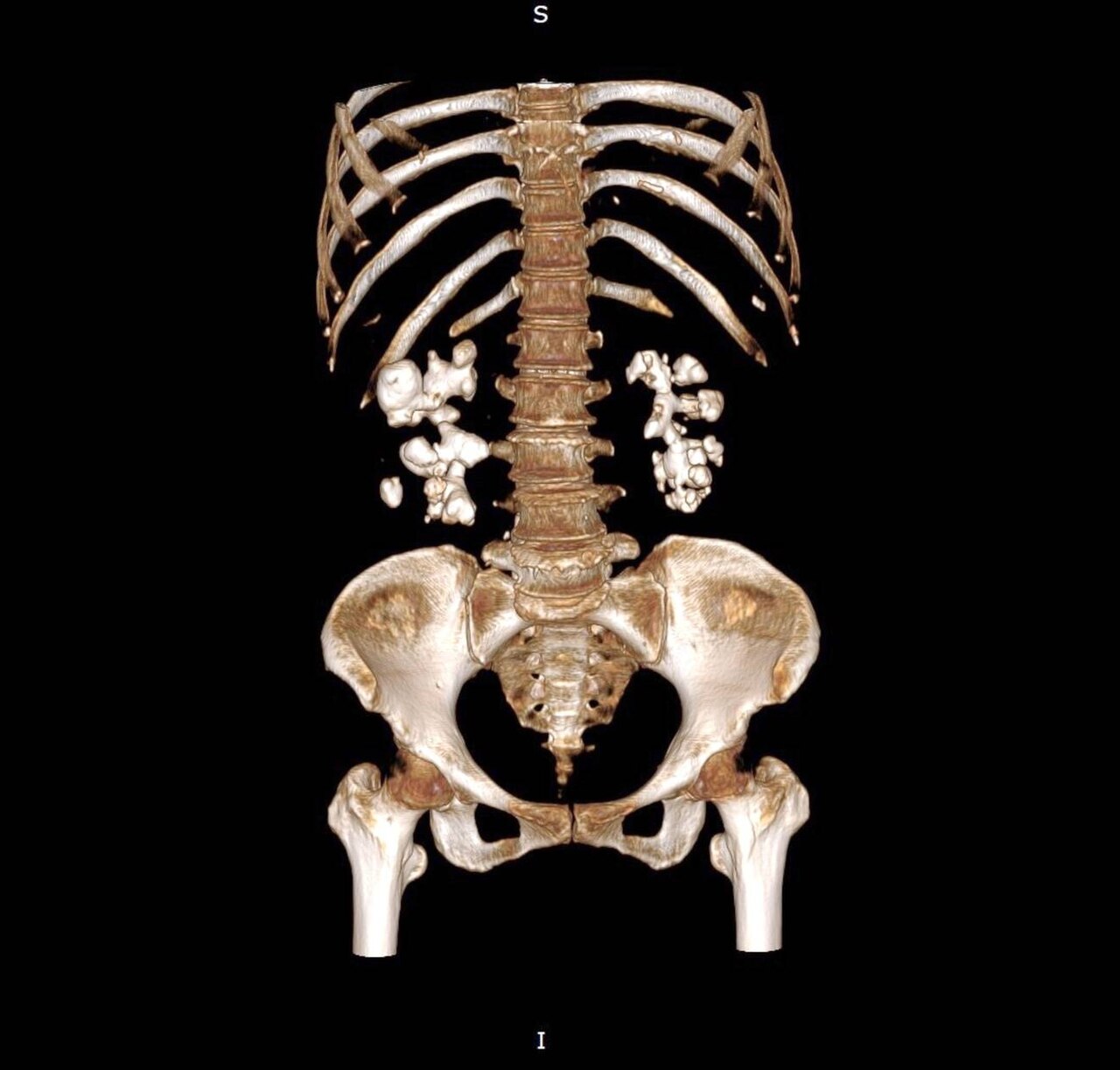
Non-contrast CT KUB (kidneys–ureters–bladder) is the imaging reference standard for suspected ureteric colic, with > 95% sensitivity. NICE NG118 (2019) recommends CT KUB within 24 hours of presentation. Low-dose protocols keep dose under 3 mSv. Ultrasound is preferred first-line in pregnancy and children.
CT KUB is a rapid, non-contrast scan protocol used specifically for suspected renal or ureteric stones. It identifies stones as small as 1 mm, distinguishes ureteric colic from important mimics (leaking AAA, appendicitis, ovarian pathology), and gives stone location + size + Hounsfield density to guide treatment. NICE NG118 recommends CT KUB within 24 hours of adult presentation with suspected renal colic. Radiation dose has fallen dramatically with iterative reconstruction and low-dose protocols — many services now perform sub-2 mSv KUBs without loss of sensitivity. Ultrasound is preferred first-line in pregnancy (any radiation avoidance), in children (Image Gently), and in known-stone follow-up. Stone size guides management: < 5 mm most pass spontaneously (medical expulsive therapy with alpha-blocker if 5–10 mm ureteric stones); > 10 mm typically need urology intervention (ureteroscopy, shockwave lithotripsy, or PCNL); infection + obstruction is a urological emergency requiring urgent decompression (JJ stent or nephrostomy).
- Radiation dose
- ~2–3 mSv (low-dose protocols)
- Sensitivity
- > 95% for ureteric stones
- Contrast
- None
- Alternative
- US in pregnancy/children
- Timing
- Within 24 h (NICE NG118)
Stone size + typical management
| Size | Typical outcome |
|---|---|
| < 5 mm ureteric | Passes spontaneously; analgesia + hydration |
| 5 – 10 mm ureteric | MET (alpha-blocker) or intervention |
| > 10 mm | Ureteroscopy / SWL / PCNL |
| Any + infection + obstruction | Emergency decompression |
CT vs ultrasound for renal colic
CT KUB > 95% sensitivity for stones; ultrasound ~40–85% (highly operator-dependent).
CT gives stone size, density (helps predict SWL success), and rules out mimics.
Ultrasound remains first-line in pregnancy and children to avoid radiation.
Ultrasound reliably shows hydronephrosis but often misses ureteric stones.
- Colic + fever + rigors — obstructed infected system; emergency admission.
- Colic + solitary kidney or AKI — urgent urology.
- Colic + pregnancy — ultrasound first, MR urography if needed.
Suspected renal colic workup
- 1Adult, non-pregnant?CT KUB within 24 h (NICE NG118).
- 2Pregnant?Ultrasound + MR urography if needed.
- 3Child?Ultrasound first.
- 4Fever + obstructed system?Emergency urology.
Related questions people ask
Frequently asked questions
- CT KUB is the reference standard for adult renal colic.
- Perform within 24 hours (NICE NG118).
- Ultrasound is preferred in pregnancy and children.
- Size and location determine management.
- Infection + obstruction is a urological emergency.
References
2 sources- NICE NG118Renal and ureteric stones
UK national guideline.
nice.org.uk
- EAUUrolithiasis Guidelines
European urology guideline.
uroweb.org
Related articles
 Imaging & Diagnostic Tests
Chest X-ray Explained: What It Shows, When You Need One, and Radiation Dose
A chest X-ray delivers a chest radiation dose equivalent to a few days of background radiation. Learn what it detects and what it misses.
 Imaging & Diagnostic Tests
CT Head Scan Explained: When It Is Needed for Stroke, Trauma and Headache
A non-contrast CT head is the first imaging test in stroke and head trauma. Learn NICE criteria, radiation dose, and what CT misses.
 Imaging & Diagnostic Tests
CTPA (CT Pulmonary Angiogram) Explained: Diagnosing Pulmonary Embolism
CTPA is the reference standard for pulmonary embolism. Learn indications, radiation dose, contrast risks, and pregnancy alternatives.