Endoscopic Ultrasound (EUS) Explained: Pancreatic Lesions, Small Stones and Staging
EUS combines endoscopy with ultrasound — invaluable for pancreatic and ampullary disease. Learn indications and FNA/FNB sampling.
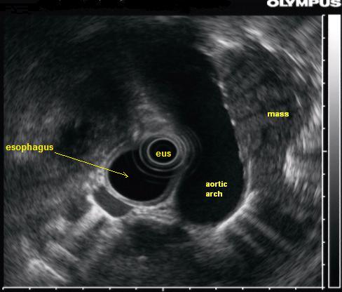
Endoscopic ultrasound (EUS) combines an endoscope with a high-frequency ultrasound transducer, giving high-resolution images from inside the gut. It excels at pancreatic and ampullary lesions, small bile-duct stones missed on MRCP, subepithelial GI tumours, mediastinal staging, and provides FNA/FNB tissue sampling.
EUS resolves lesions invisible or ambiguous on CT/MRI because of the proximity of the transducer to target structures. Radial EUS gives 360° cross-sectional views; linear EUS enables real-time needle guidance for FNA (fine-needle aspiration) or FNB (fine-needle biopsy). Standard indications: (1) pancreatic solid mass characterisation and biopsy (sensitivity for pancreatic adenocarcinoma > 90%); (2) pancreatic cystic lesions (differentiate IPMN, MCN, SCA, pseudocyst) — combined imaging + fluid analysis (CEA, amylase, glucose, molecular markers); (3) subepithelial GI lesions (GIST, leiomyoma, lipoma, ectopic pancreas) — sizing and biopsy; (4) small (< 6 mm) common bile duct stones missed by MRCP; (5) mediastinal lymph-node staging in NSCLC and oesophageal cancer; (6) rectal cancer T-staging; (7) chronic pancreatitis diagnosis (Rosemont criteria). Therapeutic EUS is expanding rapidly: EUS-guided biliary drainage in failed ERCP, cyst drainage, coeliac plexus block for pancreatic pain, gallbladder drainage. Complications rate ~1–2% (bleeding, perforation, pancreatitis after FNA).
- Sensitivity — pancreatic cancer
- > 90%
- Small CBD stone
- EUS > MRCP
- Cyst fluid analysis
- CEA, amylase, glucose, molecular
- Preferred approach for FNA/FNB
- Linear EUS
- Complication rate
- ~1–2%
EUS strengths vs alternatives
| Question | Best |
|---|---|
| Small bile-duct stone (MRCP-negative) | EUS |
| Pancreatic solid mass biopsy | EUS-FNB |
| Pancreatic cyst characterisation | EUS + cyst fluid |
| Subepithelial GI lesion | EUS |
| Mediastinal staging in NSCLC | EBUS + EUS |
When to consider EUS
Indeterminate pancreatic lesion on CT/MRI.
Pancreatic cyst characterisation.
Suspicion of small CBD stone with negative MRCP.
Subepithelial lesion needing tissue sampling.
Chronic pancreatitis diagnosis.
Mediastinal / peripancreatic staging.
- Solid pancreatic mass — MDT within 2 weeks (pancreatic cancer pathway).
- Post-EUS abdominal pain > 6 h — assess for post-FNA pancreatitis.
- Cyst with worrisome features (thick septa, mural nodule, size > 3 cm) — surgical review.
Pancreatic lesion workup
- 1Solid pancreatic lesion?CT staging + EUS-FNB.
- 2Cystic lesion?MRI + EUS + cyst fluid.
- 3MRCP-negative + strong suspicion of small CBD stone?EUS.
- 4Subepithelial GI lesion?EUS ± FNB.
Related questions people ask
Frequently asked questions
- EUS is unmatched for pancreatic and ampullary detail.
- Small CBD stones missed by MRCP are seen on EUS.
- FNB enables tissue diagnosis without open biopsy.
- Cyst fluid analysis differentiates neoplastic vs non-neoplastic.
- Therapeutic EUS is a growing field.
References
2 sources- ESGEEUS Guidelines
European endoscopy society.
esge.com
- ASGEStandards of Practice for EUS
US endoscopy society.
asge.org
Related articles
 Imaging & Diagnostic Tests
Chest X-ray Explained: What It Shows, When You Need One, and Radiation Dose
A chest X-ray delivers a chest radiation dose equivalent to a few days of background radiation. Learn what it detects and what it misses.
 Imaging & Diagnostic Tests
CT Head Scan Explained: When It Is Needed for Stroke, Trauma and Headache
A non-contrast CT head is the first imaging test in stroke and head trauma. Learn NICE criteria, radiation dose, and what CT misses.
 Imaging & Diagnostic Tests
CTPA (CT Pulmonary Angiogram) Explained: Diagnosing Pulmonary Embolism
CTPA is the reference standard for pulmonary embolism. Learn indications, radiation dose, contrast risks, and pregnancy alternatives.