MRCP Explained: Non-Invasive Imaging of Bile Ducts and Pancreas
MRCP visualises bile and pancreatic ducts without contrast or radiation. Learn indications, comparison with ERCP, and pitfalls.
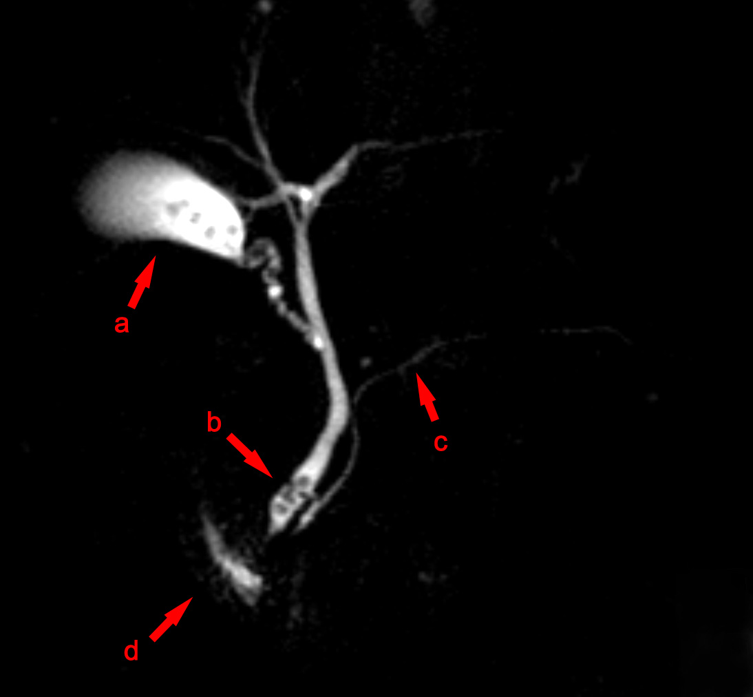
MRCP is a non-invasive MRI sequence that visualises bile ducts and pancreatic ducts without contrast or radiation. It is the first-line diagnostic test for suspected bile-duct stones (choledocholithiasis) after ultrasound and for primary sclerosing cholangitis. ERCP is now reserved for therapy — not diagnosis.
MRCP uses heavily T2-weighted MRI sequences that make static or slow-moving fluid (bile, pancreatic secretions) appear very bright, allowing detailed depiction of the pancreaticobiliary tree without contrast or radiation. Sensitivity for common bile-duct stones > 90%; slightly reduced for stones < 6 mm. Key indications: suspected choledocholithiasis after ultrasound; unexplained biliary dilatation; primary sclerosing cholangitis (beaded strictures); primary biliary cholangitis; chronic pancreatitis (ductal changes); IPMN and pancreatic cystic lesion characterisation; congenital biliary anomalies. MRCP has largely replaced diagnostic ERCP because ERCP carries a 3–5% pancreatitis risk. Endoscopic ultrasound (EUS) is superior for small stones near the ampulla and for characterising pancreatic solid lesions. Secretin-enhanced MRCP improves visualisation of pancreatic ducts and may help detect chronic pancreatitis. Interpretation: normal common bile duct diameter is ≤ 6 mm (up to 8 mm post-cholecystectomy or age > 60); pancreatic duct ≤ 3 mm.
- Radiation
- None
- Contrast
- Usually none
- Sensitivity — CBD stones
- > 90%
- Now first-line over
- Diagnostic ERCP
- Alternative
- EUS for small stones near ampulla
MRCP vs ERCP vs EUS
| Factor | MRCP | ERCP | EUS |
|---|---|---|---|
| Radiation | None | Fluoroscopic | None |
| Diagnostic | Yes | Historic | Yes |
| Therapeutic | No | Yes (stent, sphincterotomy) | FNA |
| Small stone sensitivity | Moderate | High | Highest |
| Pancreatitis risk | None | 3–5% | Very low |
When MRCP is indicated
Ultrasound shows biliary dilatation without cause.
Suspected choledocholithiasis with abnormal LFTs.
Follow-up of primary sclerosing cholangitis (annual MRCP + MDT).
Chronic pancreatitis assessment.
Pancreatic cystic lesion characterisation.
- Cholangitis (fever + jaundice + RUQ pain) — emergency, therapeutic ERCP after ultrasound/MRCP.
- MRCP suggests malignant stricture — hepatobiliary MDT.
- Rising bilirubin + weight loss — urgent imaging pathway.
Biliary workup
- 1Ultrasound shows dilated ducts?MRCP.
- 2MRCP shows stones + symptoms?Therapeutic ERCP.
- 3MRCP suggests stricture?EUS + biopsy + hepatobiliary MDT.
- 4Small stone near ampulla, MRCP negative?EUS.
Related questions people ask
Frequently asked questions
- MRCP is the first-line non-invasive biliary/pancreatic duct imaging.
- It has replaced diagnostic ERCP for most indications.
- EUS is complementary for small ampullary stones.
- Cholangitis needs therapeutic ERCP after diagnostic imaging.
- PSC requires annual MRCP surveillance.
References
2 sources- ESGEEndoscopic Management of Choledocholithiasis Guidelines
European endoscopy society.
esge.com
- BSGPrimary Sclerosing Cholangitis Guidelines
British Society of Gastroenterology.
gut.bmj.com
Related articles
 Imaging & Diagnostic Tests
Chest X-ray Explained: What It Shows, When You Need One, and Radiation Dose
A chest X-ray delivers a chest radiation dose equivalent to a few days of background radiation. Learn what it detects and what it misses.
 Imaging & Diagnostic Tests
CT Head Scan Explained: When It Is Needed for Stroke, Trauma and Headache
A non-contrast CT head is the first imaging test in stroke and head trauma. Learn NICE criteria, radiation dose, and what CT misses.
 Imaging & Diagnostic Tests
CTPA (CT Pulmonary Angiogram) Explained: Diagnosing Pulmonary Embolism
CTPA is the reference standard for pulmonary embolism. Learn indications, radiation dose, contrast risks, and pregnancy alternatives.