ERCP Explained: Therapeutic Endoscopic Biliary and Pancreatic Intervention
ERCP is now a therapeutic tool for bile-duct stones, strictures, and leaks. Learn its risks, pancreatitis mitigation, and modern indications.
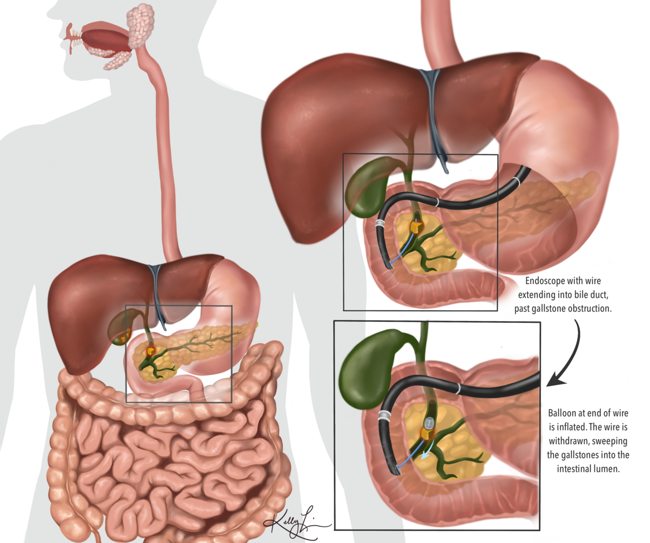
ERCP is now primarily a therapeutic procedure — diagnostic ERCP has been replaced by MRCP and EUS. Indications: bile-duct stone extraction, biliary drainage in obstructive jaundice, stenting of malignant strictures, treatment of bile leaks, sphincterotomy for sphincter of Oddi dysfunction. Post-ERCP pancreatitis occurs in 3–5%; rectal indomethacin reduces this risk.
ERCP combines side-viewing duodenoscopy with fluoroscopic contrast injection of the biliary and pancreatic ducts. Once the diagnostic and therapeutic mainstay of biliary disease, it is now almost exclusively therapeutic — diagnostic imaging is done with MRCP or EUS. Key therapeutic manoeuvres: biliary sphincterotomy, balloon or basket stone extraction, plastic or metal stent insertion, biliary dilation, tissue sampling (brushings). Indications: choledocholithiasis with cholangitis or persistent obstruction; biliary drainage in obstructive jaundice (benign or malignant strictures); tissue diagnosis of hilar or distal biliary strictures; management of post-cholecystectomy bile leaks; sphincter of Oddi dysfunction (controversial); pancreatic duct interventions in chronic pancreatitis. Complications: post-ERCP pancreatitis (3–5%, severe in ~10% of cases), bleeding (~1%), perforation (~0.5%), cholangitis (~1%). Rectal indomethacin 100 mg pre-procedure reduces post-ERCP pancreatitis by ~50% (INDOMETH-RCT). Prophylactic pancreatic duct stent reduces pancreatitis in high-risk cases. ERCP now competes with EUS-guided biliary drainage in surgically altered anatomy.
- Primary role
- Therapeutic (no longer diagnostic)
- Post-ERCP pancreatitis
- 3–5% (severe ~10% of those)
- Prevention
- Rectal indomethacin 100 mg pre-procedure
- Preferred diagnostic
- MRCP or EUS first
- Sedation
- Conscious sedation or GA
When ERCP vs alternative
| Question | Best |
|---|---|
| Diagnostic biliary imaging | MRCP |
| Small ampullary stone | EUS ± ERCP |
| Bile duct stone extraction | ERCP |
| Malignant biliary stricture drainage | ERCP or EUS-guided drainage |
| Post-cholecystectomy bile leak | ERCP with stent |
Reducing post-ERCP pancreatitis
Rectal indomethacin 100 mg for all patients unless contraindicated (ESGE 2020).
Aggressive IV Ringer's lactate hydration.
Prophylactic pancreatic-duct stent in high-risk (young female, prior post-ERCP pancreatitis, sphincter of Oddi dysfunction).
Avoid difficult repeated cannulations.
- Post-ERCP abdominal pain lasting > 6 h — treat as pancreatitis.
- Fever + rigors after ERCP — cholangitis.
- Cholangitis before ERCP — urgent decompression + antibiotics.
ERCP indications
- 1Bile-duct stone + cholangitis?Urgent ERCP.
- 2Malignant biliary obstruction?ERCP stent + tissue.
- 3Post-cholecystectomy leak?ERCP with stent.
- 4Diagnostic uncertainty?MRCP or EUS first.
Related questions people ask
Frequently asked questions
- ERCP is now a therapeutic procedure.
- MRCP and EUS have replaced diagnostic ERCP.
- Rectal indomethacin reduces post-ERCP pancreatitis.
- Cholangitis requires urgent ERCP.
- Post-cholecystectomy bile leaks are treated with ERCP stents.
References
2 sources- ESGEERCP Clinical Guidelines
European endoscopy society.
esge.com
- ASGEStandards of Practice for ERCP
American Society for Gastrointestinal Endoscopy.
asge.org
Related articles
 Imaging & Diagnostic Tests
Chest X-ray Explained: What It Shows, When You Need One, and Radiation Dose
A chest X-ray delivers a chest radiation dose equivalent to a few days of background radiation. Learn what it detects and what it misses.
 Imaging & Diagnostic Tests
CT Head Scan Explained: When It Is Needed for Stroke, Trauma and Headache
A non-contrast CT head is the first imaging test in stroke and head trauma. Learn NICE criteria, radiation dose, and what CT misses.
 Imaging & Diagnostic Tests
CTPA (CT Pulmonary Angiogram) Explained: Diagnosing Pulmonary Embolism
CTPA is the reference standard for pulmonary embolism. Learn indications, radiation dose, contrast risks, and pregnancy alternatives.