HIDA Scan Explained: Hepatobiliary Scintigraphy for Cholecystitis and Bile Leaks
HIDA scan images bile flow using a radiotracer. Learn when it diagnoses acute cholecystitis, bile leaks, and biliary dyskinesia.
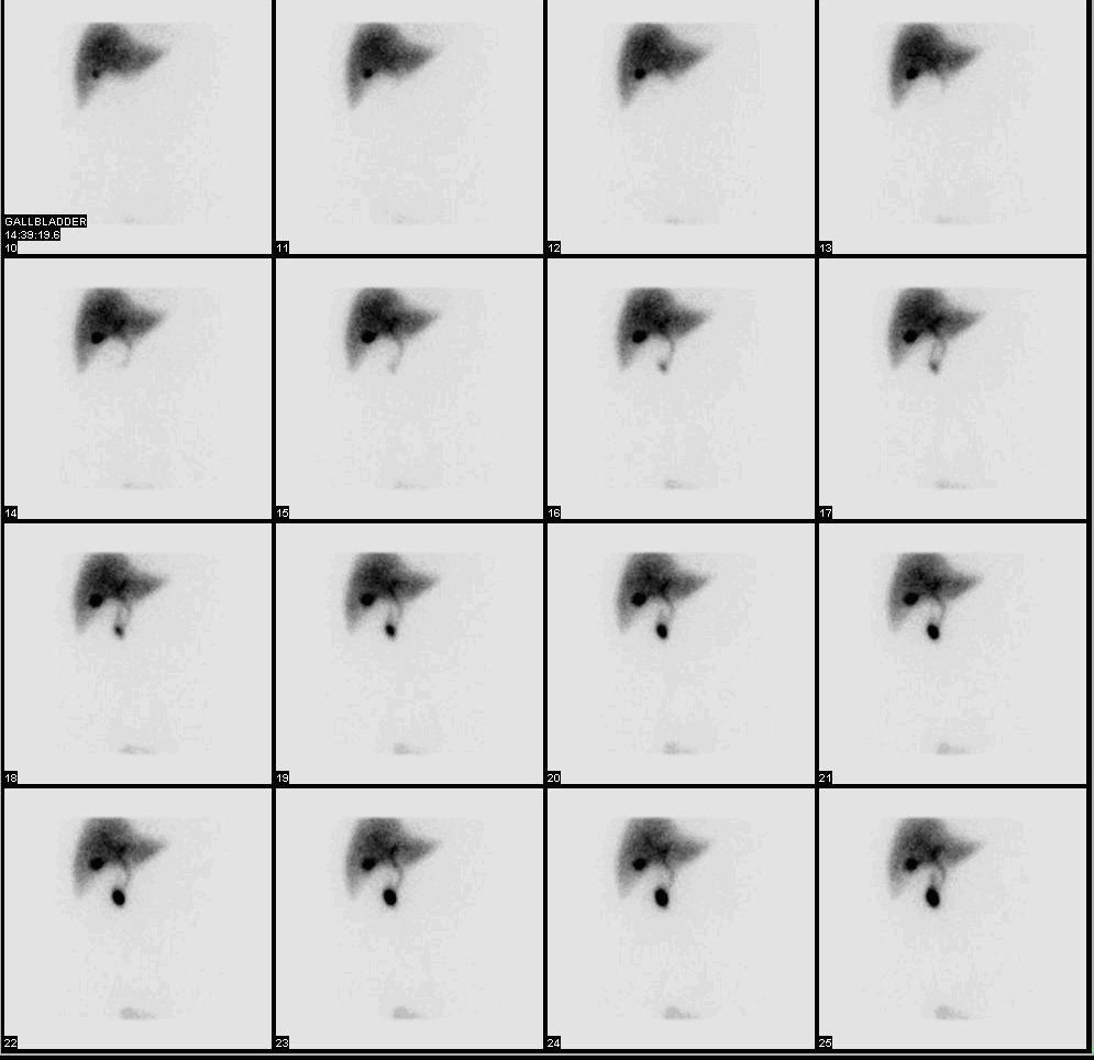
HIDA scan uses 99mTc-labelled hepatobiliary iminodiacetic acid to image bile flow through the liver, gallbladder, and bile ducts. Non-visualisation of the gallbladder at 60 minutes suggests acute cholecystitis (sensitivity ~95%). It also assesses gallbladder ejection fraction (< 35% suggests biliary dyskinesia) and detects bile leaks after cholecystectomy.
HIDA (also called cholescintigraphy) is a functional biliary test performed when ultrasound is equivocal or bile-flow physiology is being assessed. In acute cholecystitis, ultrasound typically shows gallstones, wall thickening, and pericholecystic fluid — usually sufficient. HIDA is reserved for cases where these findings are equivocal: sensitivity for acute cholecystitis > 95%, specificity > 90%. Findings: non-visualisation of the gallbladder at 60 minutes = cystic duct obstruction (acute cholecystitis); delayed visualisation at 4 hours = chronic cholecystitis. CCK-stimulated gallbladder ejection fraction (GBEF) < 35% supports biliary dyskinesia (a controversial diagnosis — Rome IV criteria emphasise functional gallbladder disorder). Post-cholecystectomy bile leak: tracer accumulates outside biliary tree. Sphincter of Oddi dysfunction: prolonged transit through common bile duct with delayed appearance in bowel. Radiation dose ~5–7 mSv. Preparation: fast 4–6 hours (empty gallbladder falsely fails to fill); avoid opioids (constrict sphincter of Oddi); recent parenteral nutrition or fasting > 24 hours may cause false non-filling (add morphine or CCK challenge).
- Radiation dose
- ~5–7 mSv
- Sensitivity — acute cholecystitis
- > 95%
- GBEF cutoff
- < 35% supports dyskinesia
- Preparation
- Fast 4–6 h; avoid opioids
- Alternative
- Ultrasound first-line
HIDA findings + interpretation
| Finding | Meaning |
|---|---|
| Gallbladder not visualised at 60 min | Acute cholecystitis |
| Delayed at 4 h | Chronic cholecystitis |
| GBEF < 35% | Biliary dyskinesia |
| Extraluminal tracer | Bile leak |
| Prolonged CBD transit | Sphincter of Oddi dysfunction |
When HIDA is chosen
Equivocal ultrasound with high clinical suspicion of cholecystitis.
Suspected chronic acalculous gallbladder disease (biliary dyskinesia).
Post-cholecystectomy bile leak evaluation.
Sphincter of Oddi dysfunction assessment.
Biliary atresia in neonates (paediatric practice).
- Acute cholecystitis with sepsis — early cholecystectomy.
- Bile leak post-cholecystectomy — MDT with hepatobiliary surgery.
- Neonatal jaundice + biliary atresia suspicion — urgent workup (Kasai procedure).
RUQ pain workup — HIDA role
- 1US equivocal for cholecystitis?HIDA.
- 2Chronic biliary symptoms + normal US?HIDA with CCK for GBEF.
- 3Post-op bile leak?HIDA.
Related questions people ask
Frequently asked questions
- HIDA is a functional biliary test.
- Non-visualised gallbladder at 60 min = cholecystitis.
- GBEF < 35% supports biliary dyskinesia.
- Excellent for bile leaks after cholecystectomy.
- Ultrasound remains first-line for gallbladder disease.
References
2 sources- SNMMI/EANMHIDA Procedure Guidelines
Nuclear medicine practice guideline.
snmmi.org
- Tokyo Guidelines 2018Acute Cholecystitis
International cholecystitis criteria.
onlinelibrary.wiley.com
Related articles
 Imaging & Diagnostic Tests
Chest X-ray Explained: What It Shows, When You Need One, and Radiation Dose
A chest X-ray delivers a chest radiation dose equivalent to a few days of background radiation. Learn what it detects and what it misses.
 Imaging & Diagnostic Tests
CT Head Scan Explained: When It Is Needed for Stroke, Trauma and Headache
A non-contrast CT head is the first imaging test in stroke and head trauma. Learn NICE criteria, radiation dose, and what CT misses.
 Imaging & Diagnostic Tests
CTPA (CT Pulmonary Angiogram) Explained: Diagnosing Pulmonary Embolism
CTPA is the reference standard for pulmonary embolism. Learn indications, radiation dose, contrast risks, and pregnancy alternatives.