Exercise Stress Test Explained: Bruce Protocol, ST Changes and When to Use It
The treadmill ECG is falling out of favour but still has a role. Learn the Bruce protocol, ST criteria, and when CCTA or stress imaging is preferred.
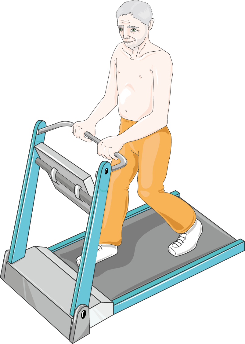
The exercise stress test (treadmill ECG) records the ECG during graded exercise (Bruce protocol) looking for ST-segment changes suggesting inducible ischaemia. Sensitivity is only ~68% and specificity ~77% — leading NICE to remove it as first-line for stable chest pain in favour of CCTA (CG95). It remains useful for functional capacity, arrhythmia unmasking, and exercise-related symptoms.
The exercise ECG was the historic workhorse of ischaemic heart disease diagnosis but modern imaging has largely displaced it. NICE CG95 (2016 update) removed exercise ECG as a first-line diagnostic test for stable chest pain, recommending CCTA instead. Its remaining roles: (1) risk stratification in known coronary disease; (2) exercise-related symptoms (syncope, arrhythmia); (3) functional capacity for cardiac rehabilitation planning; (4) low-cost triage where CCTA/stress imaging unavailable. Bruce protocol: 7 three-minute stages of increasing speed and incline. Positive result: ≥ 1 mm horizontal or downsloping ST depression 80 ms after J point, in ≥ 2 contiguous leads. Higher-risk features (Duke Treadmill Score): early positive at low workload, ST elevation without infarct, hypotensive response, ventricular arrhythmia, inability to reach 85% predicted maximum heart rate. Stress imaging (stress echo or MPI) has better sensitivity/specificity and can be used when exercise ECG is uninterpretable (baseline LBBB, pacing, digoxin, LVH) or the patient cannot exercise (pharmacological stress).
- Protocol
- Bruce (or modified Bruce)
- Positive ST change
- ≥ 1 mm horizontal/downsloping depression
- Sensitivity
- ~68%
- Uninterpretable
- LBBB, pacing, LVH, digoxin
- Preferred alternative
- CCTA or stress imaging
Stress-test choices
| Situation | Preferred test |
|---|---|
| Suspected stable angina (new) | CCTA (NICE first-line) |
| Known CAD, functional stratification | Exercise stress or stress echo |
| Cannot exercise | Pharmacological stress (dobutamine echo or vasodilator MPI) |
| Uninterpretable baseline ECG | Stress imaging |
| Exercise-related symptoms | Exercise stress test |
Preparation + safety
Fast 2 h before; avoid caffeine 12 h.
Continue beta-blocker if on for arrhythmia; discuss with cardiologist otherwise.
Wear comfortable footwear; report chest discomfort to the supervising clinician.
Absolute contraindications: acute MI within 2 days, unstable angina, uncontrolled arrhythmia, active endocarditis, aortic dissection, acute PE.
- Chest pain at rest — not for stress test; treat as ACS.
- Positive stress test with early ischaemia — urgent cardiology.
- Syncope + exertion — full cardiac workup before stress testing.
Suspected angina pathway
- 1New chest pain, no known CAD?CCTA (NICE first-line).
- 2Known CAD, needs functional data?Exercise stress or stress echo.
- 3Cannot exercise or uninterpretable baseline?Pharmacological stress imaging.
Related questions people ask
Frequently asked questions
- Exercise stress ECG is no longer first-line for suspected angina (NICE).
- Bruce protocol looks for ≥ 1 mm ST changes.
- Sensitivity is modest; imaging alternatives are better.
- Useful for functional capacity and exercise-related symptoms.
- Uninterpretable baseline needs stress imaging.
References
2 sources- NICE CG95Chest pain of recent onset
UK national guideline.
nice.org.uk
- ACC/AHAExercise Testing Guideline
US cardiology.
jacc.org
Related articles
 Imaging & Diagnostic Tests
Chest X-ray Explained: What It Shows, When You Need One, and Radiation Dose
A chest X-ray delivers a chest radiation dose equivalent to a few days of background radiation. Learn what it detects and what it misses.
 Imaging & Diagnostic Tests
CT Head Scan Explained: When It Is Needed for Stroke, Trauma and Headache
A non-contrast CT head is the first imaging test in stroke and head trauma. Learn NICE criteria, radiation dose, and what CT misses.
 Imaging & Diagnostic Tests
CTPA (CT Pulmonary Angiogram) Explained: Diagnosing Pulmonary Embolism
CTPA is the reference standard for pulmonary embolism. Learn indications, radiation dose, contrast risks, and pregnancy alternatives.