FibroScan (Transient Elastography) Explained: Liver Fibrosis and Steatosis
FibroScan is a painless, radiation-free test that quantifies liver fibrosis and fat. Learn how it replaces most liver biopsies in NAFLD.
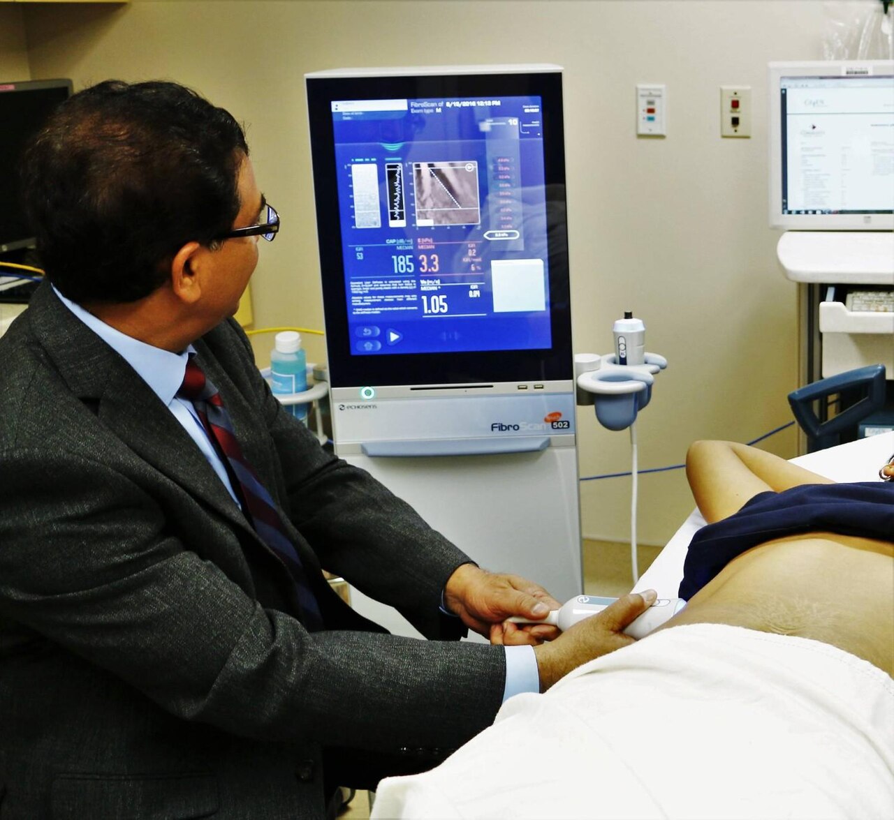
FibroScan uses transient elastography to measure liver stiffness (kPa) — a proxy for fibrosis — and CAP (controlled attenuation parameter, dB/m) — a proxy for steatosis. It has largely replaced diagnostic liver biopsy for NAFLD/MASLD staging. Values > 12 kPa suggest advanced fibrosis (F3–F4).
FibroScan (Echosens) is the most widely used point-of-care technique for non-invasive liver fibrosis assessment. It uses a low-frequency mechanical impulse and ultrasound to measure shear-wave velocity, giving a liver stiffness measurement (LSM) in kPa. The 2024 EASL–EASD–EASO MASLD clinical practice guideline endorses two-step pathways: (1) FIB-4 (age, ALT, AST, platelets) as first screen; if FIB-4 > 1.3 (or > 2 in age > 65), (2) FibroScan for confirmatory staging. Thresholds vary by aetiology: in MASLD, LSM < 8 kPa excludes advanced fibrosis with high NPV; > 12 kPa suggests F3–F4 (advanced); > 20 kPa suggests clinically significant portal hypertension. In hepatitis C, thresholds are 7 kPa (F2), 9.5 kPa (F3), 12.5 kPa (F4). CAP (dB/m) grades steatosis: > 248 mild, > 268 moderate, > 280 severe. Limitations: obesity (XL probe now standard), acute inflammation, cholestasis, congestive hepatopathy, and recent food all elevate LSM falsely — fast 3 hours before scan. Non-obese patients with borderline results may need MR elastography or liver biopsy for definitive staging.
- Radiation
- None
- LSM < 8 kPa
- Excludes advanced fibrosis (MASLD)
- LSM > 12 kPa
- Suggests F3–F4
- CAP
- Steatosis score
- Preparation
- Fast 3 h before scan
MASLD FibroScan interpretation
| LSM (kPa) | Interpretation | Action |
|---|---|---|
| < 8 | No/low fibrosis | Lifestyle; recheck 3 y |
| 8 – 12 | Indeterminate | Repeat or MR elastography |
| 12 – 20 | Advanced fibrosis (F3) | Hepatology referral |
| > 20 | Cirrhosis + portal HT | Variceal + HCC surveillance |
When to use FibroScan
Elevated ALT/AST or fatty liver on ultrasound — for staging fibrosis.
Chronic hepatitis B or C.
Alcohol-related liver disease.
Monitoring after DAA therapy in HCV.
Surveillance of MASLD to detect progression.
- LSM > 20 kPa — variceal screening + HCC surveillance.
- New ascites or jaundice — hepatology admission.
- Rising LSM on serial scans — accelerate work-up.
MASLD workup pathway
- 1Metabolic risk + raised ALT?FIB-4 first.
- 2FIB-4 > 1.3?FibroScan.
- 3LSM > 12 kPa?Hepatology.
- 4Cirrhosis?Variceal + HCC surveillance every 6 months.
Related questions people ask
Frequently asked questions
- FibroScan quantifies fibrosis and steatosis non-invasively.
- Use FIB-4 first — then FibroScan for confirmation.
- LSM > 12 kPa suggests advanced MASLD fibrosis.
- Fast 3 hours before to avoid false elevation.
- Cirrhosis triggers variceal + HCC surveillance.
References
2 sources- EASL–EASD–EASOMASLD Clinical Practice Guidelines (2024)
European joint guideline.
journal-of-hepatology.eu
- AASLDNAFLD Practice Guidance (2023)
US liver association.
aasld.org
Related articles
 Imaging & Diagnostic Tests
Chest X-ray Explained: What It Shows, When You Need One, and Radiation Dose
A chest X-ray delivers a chest radiation dose equivalent to a few days of background radiation. Learn what it detects and what it misses.
 Imaging & Diagnostic Tests
CT Head Scan Explained: When It Is Needed for Stroke, Trauma and Headache
A non-contrast CT head is the first imaging test in stroke and head trauma. Learn NICE criteria, radiation dose, and what CT misses.
 Imaging & Diagnostic Tests
CTPA (CT Pulmonary Angiogram) Explained: Diagnosing Pulmonary Embolism
CTPA is the reference standard for pulmonary embolism. Learn indications, radiation dose, contrast risks, and pregnancy alternatives.