MR Enterography (MRE) Explained: Small Bowel Crohn Disease Assessment
MR enterography is the imaging reference standard for Crohn disease. Learn how it maps activity, strictures and fistulae without radiation.
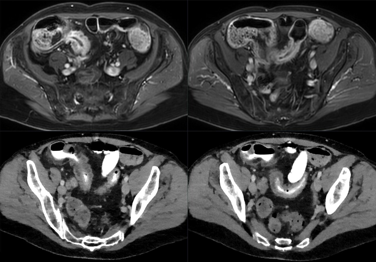
MR enterography (MRE) is the imaging reference standard for Crohn disease. It maps small-bowel inflammation, strictures, fistulae and abscesses without radiation. NICE recommends MRE for baseline staging, complication assessment, and monitoring in Crohn disease. Sensitivity for active inflammation > 90% in the terminal ileum.
MR enterography combines high-resolution abdominal MRI with oral bowel-distending contrast (usually polyethylene glycol or mannitol) to visualise the small bowel. It replaces CT enterography in most non-emergent settings due to lack of radiation — important in young IBD patients with lifetime cumulative dose concerns. Standard indications: baseline Crohn staging (extent, strictures, fistulae, abscesses); response to biologic therapy (repeat MRE 6–12 months); suspected complication (obstruction, penetrating disease); pre-surgical mapping. Activity scoring: MaRIA (Magnetic Resonance Index of Activity) and Nancy scores quantify segmental inflammation. Findings: wall thickening (> 3 mm), mural hyperenhancement, wall T2 hyperintensity, comb sign (increased vasa recta), mesenteric fat stranding, ulceration, strictures, fistulae. Preparation: fast 4 hours, then drink ~1.5 L of oral contrast over 60 minutes to distend the bowel. IV gadolinium is standard for enhancement; buscopan (hyoscine) reduces peristalsis. Compared with capsule endoscopy, MRE better detects deep transmural disease and complications but is less sensitive for mucosal-only involvement. Colonoscopy remains the reference for ileocolonic disease.
- Radiation
- None
- Sensitivity — terminal ileum activity
- > 90%
- Scoring systems
- MaRIA + Nancy
- Preparation
- Fast 4 h + oral contrast 1.5 L
- IV agents
- Gadolinium + buscopan
MRE vs alternatives in Crohn disease
| Question | Best test |
|---|---|
| Terminal ileum activity | MRE |
| Colonic disease | Colonoscopy |
| Mucosal-only involvement | Capsule endoscopy |
| Perianal fistula | Pelvic MRI |
| Acute obstruction/abscess | CT with contrast |
What findings mean
Wall thickening + hyperenhancement + T2 hyperintensity = active inflammation.
Strictured segment with prestenotic dilatation = fibrostenotic disease.
Sinus tract or fistula = penetrating disease.
Fluid collection with rim enhancement = abscess.
- Crohn + fever + tender mass — abscess suspicion.
- Recurrent obstruction — surgical review.
- Complex perianal disease — colorectal MDT.
Crohn imaging pathway
- 1New diagnosis?MRE for baseline staging.
- 2On biologic therapy?Repeat MRE 6–12 months.
- 3Acute severe symptoms?CT for emergency assessment.
- 4Perianal disease?Pelvic MRI.
Related questions people ask
Frequently asked questions
- MRE is first-line for Crohn small-bowel imaging.
- No radiation — ideal for young IBD patients.
- Detects activity, strictures, fistulae, abscesses.
- MaRIA / Nancy scores quantify activity.
- Complements colonoscopy and capsule endoscopy.
References
2 sources- ECCOGuidelines on Diagnostic Assessment in IBD
European Crohn's and Colitis Organisation.
ecco-ibd.eu
- ACR Appropriateness CriteriaSuspected Crohn Disease
US imaging appropriateness.
acr.org
Related articles
 Imaging & Diagnostic Tests
Chest X-ray Explained: What It Shows, When You Need One, and Radiation Dose
A chest X-ray delivers a chest radiation dose equivalent to a few days of background radiation. Learn what it detects and what it misses.
 Imaging & Diagnostic Tests
CT Head Scan Explained: When It Is Needed for Stroke, Trauma and Headache
A non-contrast CT head is the first imaging test in stroke and head trauma. Learn NICE criteria, radiation dose, and what CT misses.
 Imaging & Diagnostic Tests
CTPA (CT Pulmonary Angiogram) Explained: Diagnosing Pulmonary Embolism
CTPA is the reference standard for pulmonary embolism. Learn indications, radiation dose, contrast risks, and pregnancy alternatives.