MRI Knee Explained: Meniscal Tears, ACL Injury and Cartilage Assessment
MRI is the gold standard for internal knee injuries. Learn when it changes management and when clinical exam + physiotherapy are enough.
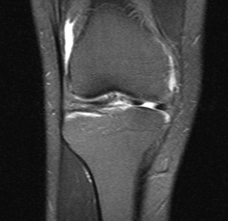
MRI knee is the imaging gold standard for meniscal tears, ACL/PCL ruptures, cartilage lesions, and bone bruising. It is not needed for every knee pain — clinical examination is highly accurate for ACL and meniscal injury when performed by experienced clinicians. MRI is justified when the result will change management (surgery or targeted rehab).
MRI knee delivers unparalleled soft-tissue contrast without radiation, resolving cruciate ligaments, menisci, cartilage, and bone marrow oedema. Sensitivity: meniscal tear 90–95%, ACL 90–98%, PCL 85–100%, cartilage lesions 70–90% (dependent on sequence). Order MRI when a positive result will change management: suspected ACL rupture in an athlete (surgery planning); locked knee (bucket-handle meniscus needing arthroscopy); rapid effusion after twist (haemarthrosis suggests ACL/fracture); failed 6 weeks of physiotherapy; suspicion of infection or osteonecrosis. Degenerative meniscal tears are common in adults over 40 — arthroscopic partial meniscectomy provides NO benefit over placebo in these patients (multiple sham-controlled RCTs). Ottawa Knee Rules guide when X-ray is needed for fracture after acute injury.
- Radiation
- None
- Sensitivity — meniscal tear
- 90–95%
- Sensitivity — ACL
- 90–98%
- Not required for
- Most degenerative knee pain > 40 y
- Acute fracture screen
- X-ray via Ottawa Knee Rules
Common MRI knee findings
| Finding | Interpretation |
|---|---|
| Complete ACL fibre disruption | ACL rupture — surgical opinion if active/young |
| Bucket-handle meniscal tear + locked knee | Arthroscopy |
| Small radial meniscal tear + age > 50 | Conservative — surgery no benefit |
| Bone marrow oedema pattern (kissing contusion) | ACL rupture pattern |
| Full-thickness cartilage defect | Cartilage repair options depending on size |
When to order MRI knee
Acute knee injury + haemarthrosis or positive Lachman/pivot shift.
Mechanical locking, clicking, or giving-way persisting despite physiotherapy.
Failed 6 weeks of conservative management + surgical opinion likely.
Suspected osteochondral fracture, osteonecrosis, or septic joint (with contrast).
- Locked knee — orthopaedic assessment for bucket-handle meniscus.
- Hot, painful, swollen joint + fever — septic arthritis, urgent aspiration.
- Unable to weight-bear after twist injury — Ottawa rules for X-ray, urgent assessment.
Knee pain workup
- 1Acute injury?Ottawa Knee Rules → X-ray if positive; MRI if surgical concern.
- 2Chronic pain > 6 weeks despite physio?MRI if surgery being considered.
- 3Suspected inflammation?ESR + CRP + aspiration; MRI with contrast if osteomyelitis suspected.
Related questions people ask
Frequently asked questions
- MRI knee is gold standard for internal injuries.
- Order when a positive result will change management.
- Arthroscopic surgery does not help degenerative meniscal tears.
- Ottawa Knee Rules screen fracture with X-ray after acute injury.
- Try 6 weeks of physio first for most chronic pain.
References
2 sources- NICEKnee pain: assessment
UK primary-care guidance.
cks.nice.org.uk
- ACR Appropriateness CriteriaAcute Trauma to the Knee
US imaging appropriateness.
acr.org
Related articles
 Imaging & Diagnostic Tests
Chest X-ray Explained: What It Shows, When You Need One, and Radiation Dose
A chest X-ray delivers a chest radiation dose equivalent to a few days of background radiation. Learn what it detects and what it misses.
 Imaging & Diagnostic Tests
CT Head Scan Explained: When It Is Needed for Stroke, Trauma and Headache
A non-contrast CT head is the first imaging test in stroke and head trauma. Learn NICE criteria, radiation dose, and what CT misses.
 Imaging & Diagnostic Tests
CTPA (CT Pulmonary Angiogram) Explained: Diagnosing Pulmonary Embolism
CTPA is the reference standard for pulmonary embolism. Learn indications, radiation dose, contrast risks, and pregnancy alternatives.