Multiparametric Prostate MRI (mpMRI) Explained: PI-RADS and Biopsy Decisions
mpMRI has transformed prostate cancer diagnosis. Learn PI-RADS scoring, when to biopsy, and how it reduces unnecessary biopsies.
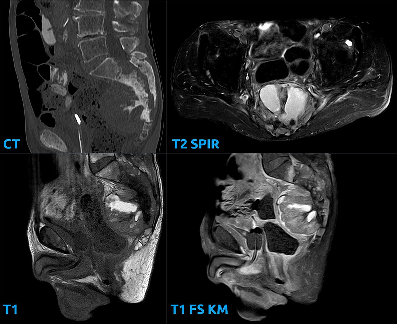
Multiparametric MRI (T2 + DWI + dynamic contrast) is now standard BEFORE prostate biopsy in men with raised PSA. NICE NG131 and EAU recommend pre-biopsy mpMRI: PI-RADS 3–5 → targeted biopsy; PI-RADS 1–2 with reassuring PSA density → biopsy may be safely deferred. The PRECISION trial showed pre-biopsy mpMRI increases detection of clinically significant cancer and reduces unnecessary biopsies.
Multiparametric prostate MRI (mpMRI) combines high-resolution T2 anatomical imaging with diffusion-weighted imaging (DWI, including apparent-diffusion-coefficient maps) and dynamic contrast-enhanced (DCE) sequences. PI-RADS v2.1 (2019, updated 2024) scores lesions 1 (very low probability of clinically significant cancer) to 5 (very high). NICE NG131 (2019, updated 2021) recommends mpMRI as the first-line investigation in men with raised PSA suitable for radical treatment — PI-RADS 3 or above triggers targeted biopsy, usually transperineal. The PRECISION trial (NEJM 2018) showed pre-biopsy mpMRI + targeted biopsy detected 38% clinically significant cancer vs 26% with systematic TRUS biopsy, and avoided biopsy in 28% of men. Transperineal biopsy has replaced TRUS in most UK/European centres due to lower sepsis rate. PSA density (PSA ÷ prostate volume) refines biopsy decisions in equivocal PI-RADS 3 lesions: > 0.15 favours biopsy. Bi-parametric MRI (T2 + DWI without contrast) is emerging as an equivalent-quality lower-cost alternative in high-throughput services.
- Radiation
- None
- Reporting
- PI-RADS v2.1
- PSA density threshold
- > 0.15 favours biopsy
- Preferred biopsy route
- Transperineal
- Key evidence
- PRECISION trial (NEJM 2018)
PI-RADS + action
| PI-RADS | Probability of clinically significant cancer | Action |
|---|---|---|
| 1 | Very low | Continue PSA surveillance |
| 2 | Low | Continue PSA surveillance |
| 3 | Intermediate | Consider biopsy — PSA density decides |
| 4 | High | Targeted biopsy |
| 5 | Very high | Targeted biopsy |
The pre-biopsy mpMRI pathway
Raised PSA (context-appropriate cut-off) + suitable for radical treatment → mpMRI.
PI-RADS 3–5 → transperineal targeted biopsy.
PI-RADS 1–2 + low PSA density → active PSA surveillance.
Suspicious mpMRI in a man on active surveillance → repeat targeted biopsy.
- PI-RADS 4/5 lesion — urgent 2-week referral to urology.
- Rising PSA + bone pain — bone imaging (bone scan / PSMA-PET).
- Post-biopsy sepsis symptoms — emergency admission.
Raised PSA workup
- 1Raised PSA + suitable for radical treatment?mpMRI.
- 2PI-RADS 4 or 5?Transperineal targeted biopsy.
- 3PI-RADS 3?PSA density decides.
- 4PI-RADS 1–2 with low density?PSA surveillance.
Related questions people ask
Frequently asked questions
- mpMRI is standard before prostate biopsy.
- PI-RADS 3–5 triggers targeted biopsy.
- PSA density refines PI-RADS 3 decisions.
- Transperineal is the preferred biopsy route.
- Negative mpMRI can safely defer biopsy in selected men.
References
3 sources- NICE NG131Prostate cancer: diagnosis and management
UK national guideline.
nice.org.uk
- EAUProstate Cancer Guideline
European association of urology.
uroweb.org
- ACRPI-RADS v2.1
US reporting system.
acr.org
Related articles
 Imaging & Diagnostic Tests
Chest X-ray Explained: What It Shows, When You Need One, and Radiation Dose
A chest X-ray delivers a chest radiation dose equivalent to a few days of background radiation. Learn what it detects and what it misses.
 Imaging & Diagnostic Tests
CT Head Scan Explained: When It Is Needed for Stroke, Trauma and Headache
A non-contrast CT head is the first imaging test in stroke and head trauma. Learn NICE criteria, radiation dose, and what CT misses.
 Imaging & Diagnostic Tests
CTPA (CT Pulmonary Angiogram) Explained: Diagnosing Pulmonary Embolism
CTPA is the reference standard for pulmonary embolism. Learn indications, radiation dose, contrast risks, and pregnancy alternatives.