Myocardial Perfusion Imaging (SPECT) Explained: Detecting Ischaemia
Nuclear stress imaging quantifies coronary flow reserve. Learn SPECT indications, radiation dose, and PET alternatives.
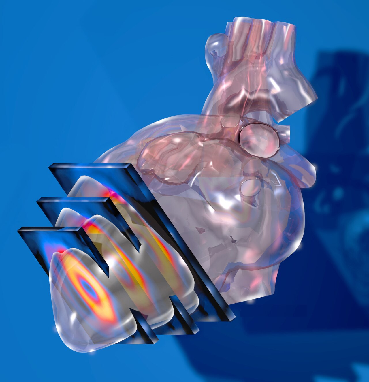
Myocardial perfusion imaging (MPI) uses SPECT with 99mTc-sestamibi (or thallium) to compare myocardial blood flow at rest and stress. Fixed defects = infarct; reversible defects = ischaemia. Sensitivity ~85%; specificity ~75%. Radiation dose 6–12 mSv. Cardiac PET (rubidium or F-18) has higher accuracy and lower dose but limited availability.
MPI SPECT remains widely used for detection of inducible ischaemia when exercise ECG or CCTA cannot answer the clinical question. Standard protocol: rest study + stress (exercise or pharmacological — dipyridamole, adenosine, regadenoson) with tracer injected at peak; comparison images demonstrate reversible defects (ischaemia), fixed defects (scar), or normal perfusion. Attenuation correction reduces false positives from breast or diaphragm. Gated SPECT adds regional wall motion + ejection fraction data. Cardiac PET (rubidium-82 or 13N-ammonia) has higher accuracy, better absolute quantification of myocardial blood flow (MBF) and coronary flow reserve, and roughly half the radiation dose — but is limited to specialist centres. Indications: intermediate pre-test probability CAD when CCTA unsuitable; risk stratification of known CAD; viability assessment (rest thallium redistribution or PET-FDG); pre-operative risk stratification before high-risk non-cardiac surgery. Downside: significant radiation dose (higher than most cardiac tests) — Choose Wisely campaigns discourage overuse in low-risk patients.
- Radiation dose
- 6–12 mSv (SPECT); 3–6 mSv (PET)
- Sensitivity
- ~85%
- Specificity
- ~75%
- Tracers
- 99mTc-sestamibi, thallium; PET: 82Rb, 13N-NH3
- PET advantage
- Absolute MBF + coronary flow reserve
MPI vs alternatives
| Situation | Best |
|---|---|
| CCTA borderline 40–70% | MPI or stress echo |
| No radiation preference | Stress echo or CMR-perfusion |
| Small woman with breast attenuation | PET or stress echo |
| Absolute MBF needed | Cardiac PET |
| Viability assessment | PET-FDG or CMR-LGE |
Interpretation basics
Reversible defect (stress worse, rest normal) = ischaemia.
Fixed defect (stress = rest, both reduced) = scar or hibernating myocardium.
Balanced ischaemia — global reduction that may falsely appear normal in three-vessel disease; PET quantifies MBF to detect this.
Transient ischaemic dilatation (TID) = high-risk marker.
- Large reversible defect + high TID — urgent cardiology.
- Post-CABG symptomatic + reversible defect — angiography.
- Recurrent symptoms + fixed defect — viability assessment before revascularisation.
When to use MPI
- 1Intermediate pre-test CAD probability + CCTA unsuitable?MPI or stress echo.
- 2Radiation concern?Prefer stress echo or CMR.
- 3Need absolute MBF?Cardiac PET.
Related questions people ask
Frequently asked questions
- MPI detects inducible ischaemia.
- Reversible defect = ischaemia; fixed = scar.
- Cardiac PET adds absolute MBF and lower dose.
- CMR and stress echo are radiation-free alternatives.
- Choose wisely — significant dose in low-risk patients.
References
2 sources- ASNC/SNMMICardiac SPECT Guidelines
US nuclear cardiology.
asnc.org
- ESC 2019 Chronic Coronary Syndromes Guideline
European cardiology.
academic.oup.com
Related articles
 Imaging & Diagnostic Tests
Chest X-ray Explained: What It Shows, When You Need One, and Radiation Dose
A chest X-ray delivers a chest radiation dose equivalent to a few days of background radiation. Learn what it detects and what it misses.
 Imaging & Diagnostic Tests
CT Head Scan Explained: When It Is Needed for Stroke, Trauma and Headache
A non-contrast CT head is the first imaging test in stroke and head trauma. Learn NICE criteria, radiation dose, and what CT misses.
 Imaging & Diagnostic Tests
CTPA (CT Pulmonary Angiogram) Explained: Diagnosing Pulmonary Embolism
CTPA is the reference standard for pulmonary embolism. Learn indications, radiation dose, contrast risks, and pregnancy alternatives.