Stress Echocardiography Explained: Exercise and Dobutamine Protocols
Stress echo combines ultrasound with exercise or dobutamine to detect inducible ischaemia. Learn indications and interpretation.
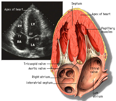
Stress echocardiography combines transthoracic echo with exercise or dobutamine to reveal inducible wall-motion abnormalities suggesting coronary artery disease. Sensitivity ~85% and specificity ~85% for significant CAD — better than treadmill ECG alone. Useful when baseline ECG is uninterpretable or when functional data are needed.
Stress echo images the left ventricle at rest and at peak stress to detect regional wall-motion abnormalities (RWMA) suggesting ischaemia. Two main protocols: (1) Exercise stress echo — treadmill or supine bike; imaged immediately post-exercise or during; preferred where possible because functional capacity and blood-pressure response add prognostic data. (2) Dobutamine stress echo — incremental IV dobutamine (10 → 40 mcg/kg/min ± atropine) to increase workload in patients who cannot exercise. Indications: suspected stable CAD when CCTA unsuitable or when downstream functional data are needed after intermediate CCTA (30–70% stenosis); pre-operative risk stratification (high-risk surgery in intermediate-risk patients); viability assessment (low-dose dobutamine + biphasic response); valve assessment under stress (low-flow low-gradient AS). Contrast enhancement (ultrasound contrast) improves endocardial border definition when > 2 segments are poorly seen. Limitations: technically demanding; poor windows in COPD/obesity. Compared with SPECT/PET perfusion, stress echo has similar accuracy and no ionising radiation, but lower spatial resolution.
- Radiation
- None
- Sensitivity
- ~85%
- Specificity
- ~85%
- Exercise vs dobutamine
- Exercise preferred when possible
- Contrast?
- Ultrasound microbubbles improve borders
Choosing a functional test
| Situation | Preferred |
|---|---|
| Suspected stable CAD, uninterpretable baseline ECG | Stress echo or MPI |
| Cannot exercise | Dobutamine stress echo or vasodilator MPI |
| CCTA borderline 40–70% | Stress echo, MPI, or FFR-CT |
| Viability assessment | Low-dose dobutamine echo or CMR-LGE |
| Low-flow low-gradient AS | Low-dose dobutamine echo |
When RWMA is positive
New or worsening wall-motion abnormality in a coronary territory.
Biphasic response (initial improvement then worsening) suggests hibernating myocardium.
Global hypokinesia suggests severe multivessel disease or non-ischaemic cardiomyopathy.
- Large territory RWMA at low workload — invasive coronary angiography.
- Chest pain + ECG changes + hypotension — stop and assess ACS.
- Sustained ventricular arrhythmia during protocol — cardiology admission.
Which stress test?
- 1Can exercise?Exercise stress echo.
- 2Cannot exercise?Dobutamine stress echo.
- 3Viability needed?Low-dose dobutamine biphasic assessment.
Related questions people ask
Frequently asked questions
- Stress echo is a validated functional ischaemia test.
- No radiation; widely available.
- Exercise is preferred; dobutamine when unable.
- Useful for viability and low-flow AS assessment.
- Complements CCTA and CMR.
References
2 sources- ASE/EACVI Stress Echo Recommendations
US/European cardiology imaging.
asecho.org
- ESC 2019 Chronic Coronary Syndromes Guideline
European cardiology.
academic.oup.com
Related articles
 Imaging & Diagnostic Tests
Chest X-ray Explained: What It Shows, When You Need One, and Radiation Dose
A chest X-ray delivers a chest radiation dose equivalent to a few days of background radiation. Learn what it detects and what it misses.
 Imaging & Diagnostic Tests
CT Head Scan Explained: When It Is Needed for Stroke, Trauma and Headache
A non-contrast CT head is the first imaging test in stroke and head trauma. Learn NICE criteria, radiation dose, and what CT misses.
 Imaging & Diagnostic Tests
CTPA (CT Pulmonary Angiogram) Explained: Diagnosing Pulmonary Embolism
CTPA is the reference standard for pulmonary embolism. Learn indications, radiation dose, contrast risks, and pregnancy alternatives.