Renal Artery Doppler Ultrasound Explained: Renovascular Hypertension Screening
Renal artery Doppler screens for renovascular disease non-invasively. Learn peak systolic velocity thresholds and when CT/MR angiography is next.
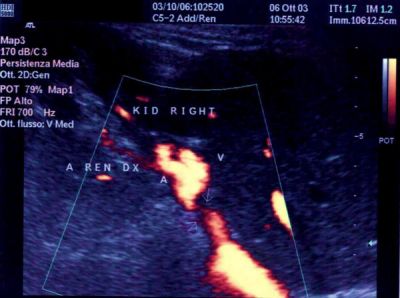
Renal artery Doppler ultrasound screens for renovascular hypertension without contrast or radiation. Peak systolic velocity (PSV) > 180 cm/s or renal-aortic ratio (RAR) > 3.5 suggests ≥ 60% stenosis. Sensitivity is operator-dependent (~85%). CT or MR angiography confirms and characterises before any revascularisation decision — although modern evidence (CORAL trial) has narrowed the indication for stenting.
Renal artery stenosis is caused mainly by atherosclerosis (older adults) or fibromuscular dysplasia (young women). Screening with Doppler ultrasound is reserved for patients with clinical clues to renovascular disease: resistant hypertension (≥ 3 drugs including a diuretic), unexplained AKI on ACEi/ARB (> 30% creatinine rise), asymmetric kidney sizes, recurrent flash pulmonary oedema. Peak systolic velocity in the renal artery > 180 cm/s (or > 200 cm/s in stricter protocols) and RAR (renal PSV / aortic PSV) > 3.5 suggest ≥ 60% stenosis. Resistive index in the renal parenchyma > 0.8 is associated with poor response to revascularisation. CT angiography or MR angiography (with caution in eGFR < 30) confirm and map. Landmark CORAL (NEJM 2014) and ASTRAL (NEJM 2009) trials showed that stenting most atherosclerotic renal artery stenoses does NOT improve outcomes over optimal medical therapy. Modern practice reserves revascularisation for: fibromuscular dysplasia (angioplasty), recurrent flash pulmonary oedema despite optimal therapy, and specific paediatric or transplant renal artery cases.
- Radiation
- None
- PSV cut-off
- > 180 cm/s
- RAR
- > 3.5
- Operator dependence
- High
- CORAL takeaway
- Stenting rarely helps atherosclerotic RAS
When to screen for renovascular disease
| Clinical clue | Screen? |
|---|---|
| Resistant hypertension (≥ 3 drugs + diuretic) | Yes |
| AKI after starting ACEi/ARB | Yes |
| Recurrent flash pulmonary oedema | Yes |
| Asymmetric kidney sizes | Yes |
| Uncomplicated hypertension | No |
When to proceed to CT/MR angiography
Positive Doppler with clinical suspicion.
Suspected fibromuscular dysplasia (young female, "string of beads" appearance).
Planning intervention — CT/MRA gives anatomic map.
Doppler technically limited (obesity, bowel gas).
- Rapid AKI on ACEi/ARB — stop drug, investigate for bilateral RAS.
- Recurrent flash pulmonary oedema — cardiology + nephrology.
- Young hypertensive female — think FMD.
Renovascular workup
- 1Clinical clues present?Doppler first.
- 2Doppler positive or clinically strong?CT or MR angiography.
- 3FMD suspected?Consider angioplasty referral.
- 4Atherosclerotic RAS?Optimal medical therapy usually.
Related questions people ask
Frequently asked questions
- Renal Doppler screens for renovascular hypertension.
- PSV > 180 cm/s + RAR > 3.5 suggests stenosis.
- CT/MR angiography confirms.
- Stenting rarely helps atherosclerotic RAS (CORAL).
- FMD may benefit from angioplasty.
References
2 sources- AHA Renal Artery Stenosis Statement
US cardiology.
ahajournals.org
- ESC 2018 Guidelines on Peripheral Arterial Diseases
European cardiology.
academic.oup.com
Related articles
 Imaging & Diagnostic Tests
Chest X-ray Explained: What It Shows, When You Need One, and Radiation Dose
A chest X-ray delivers a chest radiation dose equivalent to a few days of background radiation. Learn what it detects and what it misses.
 Imaging & Diagnostic Tests
CT Head Scan Explained: When It Is Needed for Stroke, Trauma and Headache
A non-contrast CT head is the first imaging test in stroke and head trauma. Learn NICE criteria, radiation dose, and what CT misses.
 Imaging & Diagnostic Tests
CTPA (CT Pulmonary Angiogram) Explained: Diagnosing Pulmonary Embolism
CTPA is the reference standard for pulmonary embolism. Learn indications, radiation dose, contrast risks, and pregnancy alternatives.