Reticulocyte Count: The Missing Number That Explains Every Anaemia
Reticulocytes are the bone marrow's "recent output". A high reticulocyte count in anaemia means the marrow is working; a low count means it is not. The number that most anaemia workups forget.
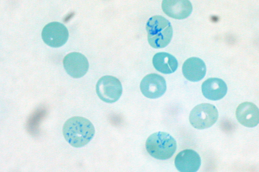
Normal reticulocyte percentage is 0.5–2.5% of red cells (absolute count 25–75 × 10⁹/L). In anaemia, this "raw" percentage is misleading because a shrunken red-cell pool makes reticulocytes look proportionally larger. Use the corrected reticulocyte count or reticulocyte production index (RPI). RPI > 2 means the marrow is responding appropriately (blood loss or haemolysis); RPI < 2 means the marrow is failing to keep up (iron, B12, folate, marrow disease, CKD).
Reticulocytes are red cells released from the bone marrow within the last 1–2 days. They mature in the bloodstream over ~24 hours before losing their reticular RNA and becoming mature erythrocytes. In anaemia, the reticulocyte count separates hypoproliferative causes (marrow underproduction) from hyperproliferative causes (blood loss, haemolysis). The corrected reticulocyte count adjusts for the patient's haematocrit: corrected retic% = observed retic% × (patient Hct ÷ normal Hct). The reticulocyte production index (RPI) further adjusts for the maturation shift — reticulocytes appear in blood earlier (up to 2.5 days) when the marrow is stressed. RPI > 2 in anaemia = appropriate response (blood loss, haemolysis, treatment response). RPI < 2 in anaemia = inappropriate response (iron/B12/folate deficiency, marrow disease, CKD-related erythropoietin deficiency).
- Normal reticulocyte %
- 0.5 – 2.5%
- Absolute reticulocyte count
- 25 – 75 × 10⁹/L
- RPI > 2 in anaemia
- Marrow responding appropriately
- RPI < 2 in anaemia
- Marrow underproduction
- Best CBC partner
- Haematocrit, MCV
Why raw reticulocyte percentage misleads
A reticulocyte % of 3% looks high — but if the patient has severe anaemia and only half the normal red-cell volume, the ABSOLUTE reticulocyte count is still normal. The correction is: corrected retic% = observed retic% × (patient Hct ÷ 45). When the marrow is stressed, it also releases reticulocytes prematurely (they normally mature 1 day in marrow, 1 in blood) — this "shift" further exaggerates the count. The RPI (reticulocyte production index) accounts for both.
A pragmatic shortcut: use the ABSOLUTE reticulocyte count (in × 10⁹/L) directly. Below ~50 × 10⁹/L in an anaemic patient is hypoproliferative; above ~100 is hyperproliferative.
Anaemia by reticulocyte response
| RPI | Category | Typical causes |
|---|---|---|
| > 2 | Hyperproliferative | Acute blood loss, haemolysis, response to iron/B12/folate/EPO |
| < 2 | Hypoproliferative | Iron deficiency, B12/folate deficiency, marrow disease, CKD, anaemia of inflammation |
When retic count changes the workup
For a normocytic anaemia (MCV 80–100), the reticulocyte count is the tie-breaker: high retic → blood loss or haemolysis; low retic → early iron deficiency, chronic disease, or renal failure. In macrocytic anaemia, high retic can point to reticulocytosis (large young cells) rather than B12/folate deficiency; check reticulocyte count before assuming B12 deficiency is the cause of a raised MCV.
After starting iron, B12, or folate replacement, expect a reticulocyte bump at 5–10 days and a rise in haemoglobin over weeks. A missing bump suggests the wrong deficiency has been treated, malabsorption, or an alternative diagnosis.
- Anaemia + very high reticulocytes + jaundice or dark urine — suspect haemolysis (check LDH, bilirubin, haptoglobin).
- Sudden reticulocytopenia after parvovirus B19 infection in sickle cell disease — aplastic crisis.
- Pancytopenia with low reticulocytes — bone marrow failure workup.
- Anaemia not responding after 4 weeks of appropriate iron replacement — reticulocyte count should have risen.
Anaemia + reticulocyte decision tree
- 1Anaemic, RPI > 2?Blood loss or haemolysis. Check LDH, haptoglobin, bilirubin. Search for GI or other bleeding site.
- 2Anaemic, RPI < 2, microcytic?Iron studies + ferritin. Iron deficiency likely.
- 3Anaemic, RPI < 2, normocytic?Ferritin, creatinine, retic, TSH. Consider anaemia of chronic disease, CKD, early iron deficiency.
- 4Anaemic, RPI < 2, macrocytic?B12, folate, TSH, reticulocyte count. Exclude alcohol, drugs, marrow disease.
- 5Anaemic on iron replacement, no rise in retic at 7–10 days?Reconsider diagnosis, check adherence, absorption; consider parenteral iron.
Related questions people ask
- What is the corrected reticulocyte count?
- How do I calculate reticulocyte production index?
- Do reticulocytes rise with iron therapy?
- How does reticulocyte count differentiate anaemias?
- What is haemolytic anaemia?
- When would reticulocytes be low in blood loss?
- Are reticulocytes elevated in B12 treatment?
Frequently asked questions
- Retic count separates hypoproliferative from hyperproliferative anaemia.
- RPI > 2 in anaemia = marrow responding; RPI < 2 = marrow failing.
- A missing retic bump at 5–10 days after treatment = wrong diagnosis or malabsorption.
- Use absolute reticulocyte count for a quick shortcut.
- Haemolysis, blood loss, and marrow recovery all raise retic.
- Iron/B12/folate deficiency, CKD, and marrow disease lower retic.
References
3 sources- ASHAnemia Guide
US haematology society educational resource.
hematology.org
- BSHAnaemia Guidelines
UK haematology society.
b-s-h.org.uk
- WHONutritional Anaemias
International anaemia framework.
who.int
Evidence Snapshot
This snapshot shows verified evidence records matched to this article. It is not a diagnosis or personal medical advice.
The reticulocyte count reports the proportion or absolute number of immature red blood cells in the blood and is used as a marker of the bone marrow's red cell production response.
Limitation. Reticulocyte percentages should be interpreted with the underlying red-cell mass, since a percentage can be misleading when total red cells are low.
In haemolytic anaemia, the reticulocyte response is typically increased as the marrow compensates for red-cell loss, whereas in marrow-suppression states the reticulocyte response is typically low despite anaemia.
Limitation. A brief lag can occur before a marrow response becomes visible, and mixed causes of anaemia can produce mixed patterns.
Related articles
 Laboratory Tests
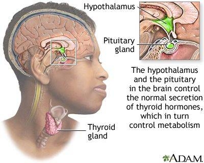
TSH Test: Normal Range, High, Low & What Your Result Actually Means
TSH is the single most important thyroid number. Understand normal ranges by age and pregnancy, what a high or low value really tells you, and the two follow-up tests that clinch the diagnosis.
 Laboratory Tests
HbA1c Test: Normal Range, Prediabetic, Diabetic Cut-offs & How to Lower It
HbA1c reflects your average blood sugar over ~3 months. Understand every threshold — normal, prediabetes, diabetes — and the realistic actions that move the number.
 Laboratory Tests
LDL Cholesterol: Optimal Numbers, High Numbers, and How to Read Yours
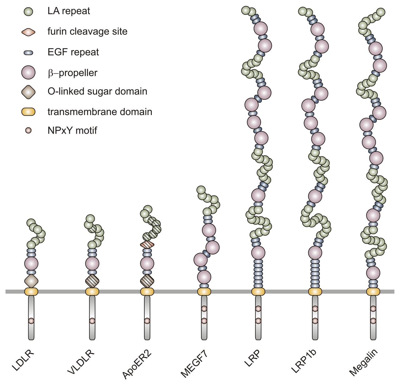
LDL is the "bad" cholesterol only if you leave the story there. Understand optimal targets by risk group, the ApoB perspective, and the interventions that actually shift the number.