Transvaginal Ultrasound Explained: Early Pregnancy, Pelvic Pain and Ovaries
Transvaginal ultrasound is more sensitive than transabdominal for pelvic structures. Learn its uses in early pregnancy, PCOS and adnexal masses.
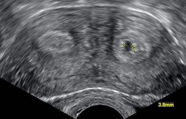
Transvaginal ultrasound (TVUS) places the probe close to the uterus and ovaries, giving much higher resolution than transabdominal scanning. It is first-line for early-pregnancy assessment, pelvic pain workup, ovarian cysts, endometrial evaluation, and PCOS ultrasound criteria.
TVUS is safe, radiation-free, and does not require a full bladder (unlike transabdominal). It is the first imaging test in most gynaecological presentations. Early pregnancy: intrauterine sac visible from ~5 weeks, fetal pole with heartbeat from ~6 weeks; empty uterus + β-hCG > 1500–2000 IU/L raises suspicion of ectopic pregnancy. PCOS ultrasound criterion (2023 international guideline) is ≥ 20 follicles per ovary OR ovarian volume ≥ 10 mL on high-resolution TVUS (AMH now an accepted alternative). Ovarian cyst characterisation follows ACR O-RADS (Ovarian-Adnexal Reporting and Data System) US v2022: categorises adnexal lesions 1 (normal) to 5 (high risk of malignancy) — CA-125 and MRI add specificity in equivocal cases. Endometrial thickness: postmenopausal bleeding + endometrial > 4 mm needs biopsy (endometrial cancer suspicion); in symptomatic women thickness < 4 mm has < 1% risk. Saline infusion sonography (SIS) improves polyp/fibroid detection. TVUS with power Doppler assesses ovarian torsion (reduced flow) but is not fully reliable — clinical suspicion + surgical intervention often needed.
- Radiation
- None
- Preparation
- Empty bladder
- Early pregnancy IUP
- Sac visible ~5 weeks
- PCOS criterion
- ≥ 20 follicles per ovary OR volume ≥ 10 mL
- Postmenopausal endometrial cut-off
- 4 mm with bleeding
When TVUS is preferred to transabdominal
| Question | Preferred |
|---|---|
| Early pregnancy assessment | TVUS |
| Adnexal mass characterisation | TVUS + O-RADS |
| Endometrial evaluation | TVUS |
| Fibroid mapping | TVUS ± SIS |
| Large mass or extreme obesity | Transabdominal + TVUS |
Key indications
Pelvic pain, bleeding, or amenorrhoea in reproductive-age women.
Suspected ectopic pregnancy — always with β-hCG.
PCOS diagnosis + workup.
Ovarian cyst characterisation with O-RADS.
Postmenopausal bleeding — endometrial assessment.
Fertility workup — antral follicle count, endometrium, uterine anomalies.
- Pelvic pain + positive pregnancy test + no intrauterine pregnancy — ectopic pathway.
- Postmenopausal bleeding — TVUS within 2 weeks.
- Ovarian mass O-RADS 4/5 — gynae-oncology MDT.
Pelvic pain workup
- 1Positive pregnancy test + pain?TVUS + β-hCG for ectopic exclusion.
- 2Adnexal mass on TVUS?O-RADS categorisation + CA-125.
- 3Postmenopausal bleeding?TVUS — biopsy if endometrium ≥ 4 mm.
Related questions people ask
Frequently asked questions
- TVUS is first-line pelvic imaging in women.
- Combine β-hCG with TVUS in suspected ectopic.
- Use O-RADS for ovarian cyst risk stratification.
- PCOS ultrasound criterion is ≥ 20 follicles or volume ≥ 10 mL.
- Postmenopausal bleeding + endometrium ≥ 4 mm → biopsy.
References
3 sources- ACRO-RADS US v2022
US reporting system.
acr.org
- International PCOS Guideline 2023
Diagnostic consensus.
monash.edu
- NICESuspected cancer: recognition and referral (NG12)
UK guideline for postmenopausal bleeding pathway.
nice.org.uk
Related articles
 Imaging & Diagnostic Tests
Chest X-ray Explained: What It Shows, When You Need One, and Radiation Dose
A chest X-ray delivers a chest radiation dose equivalent to a few days of background radiation. Learn what it detects and what it misses.
 Imaging & Diagnostic Tests
CT Head Scan Explained: When It Is Needed for Stroke, Trauma and Headache
A non-contrast CT head is the first imaging test in stroke and head trauma. Learn NICE criteria, radiation dose, and what CT misses.
 Imaging & Diagnostic Tests
CTPA (CT Pulmonary Angiogram) Explained: Diagnosing Pulmonary Embolism
CTPA is the reference standard for pulmonary embolism. Learn indications, radiation dose, contrast risks, and pregnancy alternatives.