Vestibular Testing (VNG, vHIT) Explained: Diagnosing Vertigo
Vestibular testing localises vertigo to inner ear, brain, or other systems. Learn VNG, vHIT and VEMP roles and interpretation.
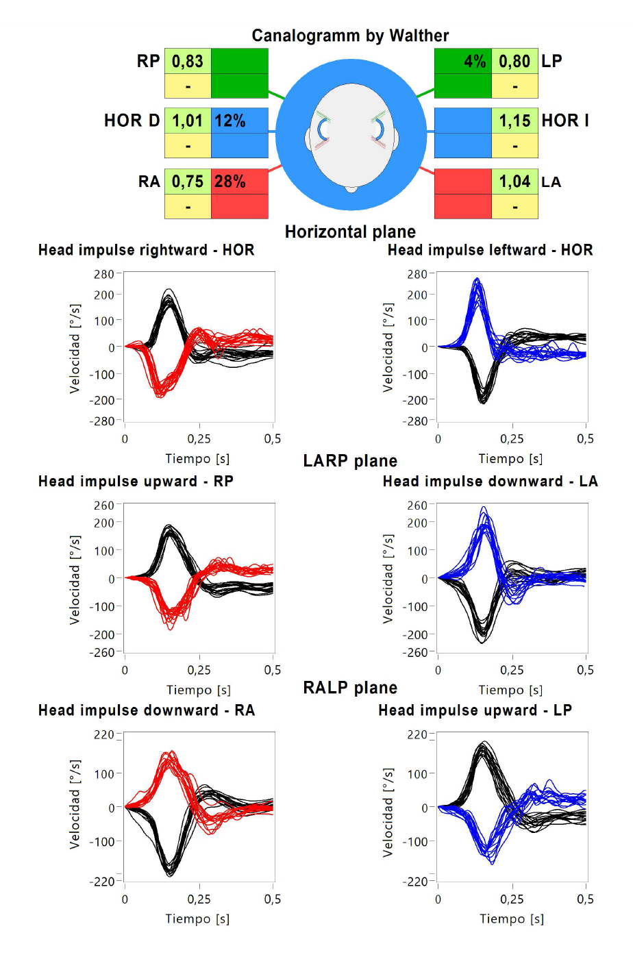
Vestibular testing localises vertigo to peripheral (inner ear) or central (brain) causes. Videonystagmography (VNG) records eye movements during positional manoeuvres, tracking, and caloric stimulation. Video head impulse test (vHIT) assesses semicircular canal function; VEMPs assess otolith function. Bedside HINTS exam distinguishes acute peripheral from central vertigo more reliably than early MRI.
Vestibular testing is a suite of physiological tests to assess balance function. VNG records eye movements via infrared goggles during: oculomotor testing (saccades, smooth pursuit, optokinetic), positional testing (Dix-Hallpike identifies BPPV), spontaneous and gaze-evoked nystagmus, and bithermal caloric stimulation (unilateral caloric weakness suggests unilateral vestibular hypofunction, e.g., vestibular neuritis). Video head impulse test (vHIT) uses high-speed cameras to detect corrective saccades during rapid head thrust — normally the eyes stay locked on target; if vestibulo-ocular reflex fails, catch-up saccades appear. Cervical (cVEMP) and ocular (oVEMP) vestibular-evoked myogenic potentials assess saccule and utricle function respectively — abnormal in Ménière and superior semicircular canal dehiscence. In ACUTE vertigo, the bedside HINTS exam (Head Impulse, Nystagmus, Test of Skew) outperforms early MRI (98% sensitivity for stroke vs 88% for MRI-DWI at < 24 h). Central signs include: direction-changing nystagmus, vertical nystagmus, skew deviation, normal head impulse test. MRI is indicated if central signs, red flags, or suspected posterior circulation stroke.
- VNG
- Records eye movements, calorics
- vHIT
- Semicircular canal function
- VEMPs
- Otolith function
- BPPV diagnosis
- Dix-Hallpike + Epley
- HINTS
- Bedside test for acute vestibular stroke
Peripheral vs central acute vertigo signs
| Sign | Peripheral | Central |
|---|---|---|
| Nystagmus direction | Unidirectional | Direction-changing or vertical |
| Head impulse test | Abnormal | Normal |
| Skew deviation | Absent | Present |
| Hearing loss | Common (labyrinthitis) | Rare |
| Neurological signs | Absent | May be present |
When to order
Recurrent or persistent vertigo unexplained by BPPV.
Suspected Ménière disease.
Post-neuritis vestibular hypofunction quantification.
Superior semicircular canal dehiscence workup.
Balance rehabilitation planning.
- Acute vertigo + central signs — stroke pathway.
- Sudden sensorineural hearing loss + vertigo — urgent ENT.
- Vertigo + severe headache + neck pain — vertebral dissection workup.
Vertigo workup
- 1Positional + < 60 s + Dix-Hallpike positive?BPPV — Epley manoeuvre.
- 2Acute persistent + normal HINTS?Vestibular neuritis.
- 3Central signs on HINTS?MRI brain + neurology.
- 4Recurrent + hearing loss + tinnitus?Consider Ménière — VEMP + VNG.
Related questions people ask
Frequently asked questions
- VNG, vHIT and VEMPs localise vestibular dysfunction.
- HINTS outperforms early MRI in acute vertigo.
- BPPV is diagnosed by Dix-Hallpike and treated by Epley.
- Central signs mandate MRI.
- Vestibular rehab improves chronic symptoms.
References
2 sources- AAO-HNS Clinical Practice Guideline: BPPV (2017 update)
US ENT society.
journals.sagepub.com
- Barany SocietyDiagnostic criteria for vestibular disorders
International vestibular society.
thejvr.com
Related articles
 Imaging & Diagnostic Tests
Chest X-ray Explained: What It Shows, When You Need One, and Radiation Dose
A chest X-ray delivers a chest radiation dose equivalent to a few days of background radiation. Learn what it detects and what it misses.
 Imaging & Diagnostic Tests
CT Head Scan Explained: When It Is Needed for Stroke, Trauma and Headache
A non-contrast CT head is the first imaging test in stroke and head trauma. Learn NICE criteria, radiation dose, and what CT misses.
 Imaging & Diagnostic Tests
CTPA (CT Pulmonary Angiogram) Explained: Diagnosing Pulmonary Embolism
CTPA is the reference standard for pulmonary embolism. Learn indications, radiation dose, contrast risks, and pregnancy alternatives.